
|
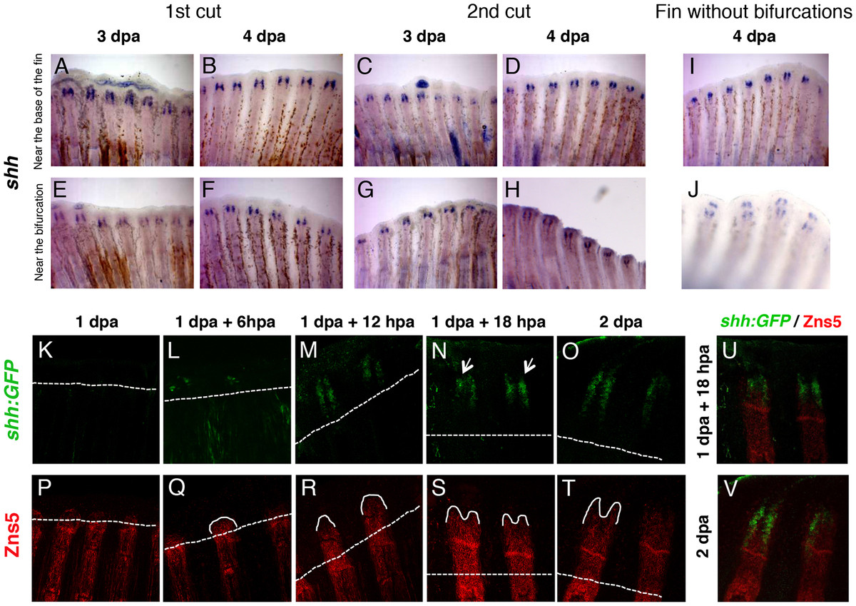
Fig. 3 The expression pattern ofshhduring regeneration does not change with the proximal-distal level or the number of amputations.(A-D) Caudal fins were amputated at 4 segments from the base of the fin (near the base of the fin) once or twice and shh expression was determined at 3 or 4 days following the amputation. (E-H) Caudal fins were amputated at 1 segment below the bifurcation (near the bifurcation) once or twice and shh expression was determined at 3 or 4 days following the amputation. (I) Caudal fins with no bifurcations were amputated near the base of the fin and shh expression was examined at 4 days following the amputation. (J) Top view of the caudal fin shown in I. (K-V) Caudal fins of 2.2shh:gfp:ABC#15 transgenic fish were amputated near the bifurcation and analyzed at different time-points after amputation by a double immunostaining with anti-GFP (green) (K-O) and anti-Zns5 (red) (P-T) antibodies. (U,V) merge of 1 dpa + 18 hpa (U) and 2 dpa (V). Dashed line represents amputation plane. hpa: hours-post-amputation; dpa: days-post-amputation. Between the first and the second amputation, fins were allowed to regenerate for 2 weeks.