
|
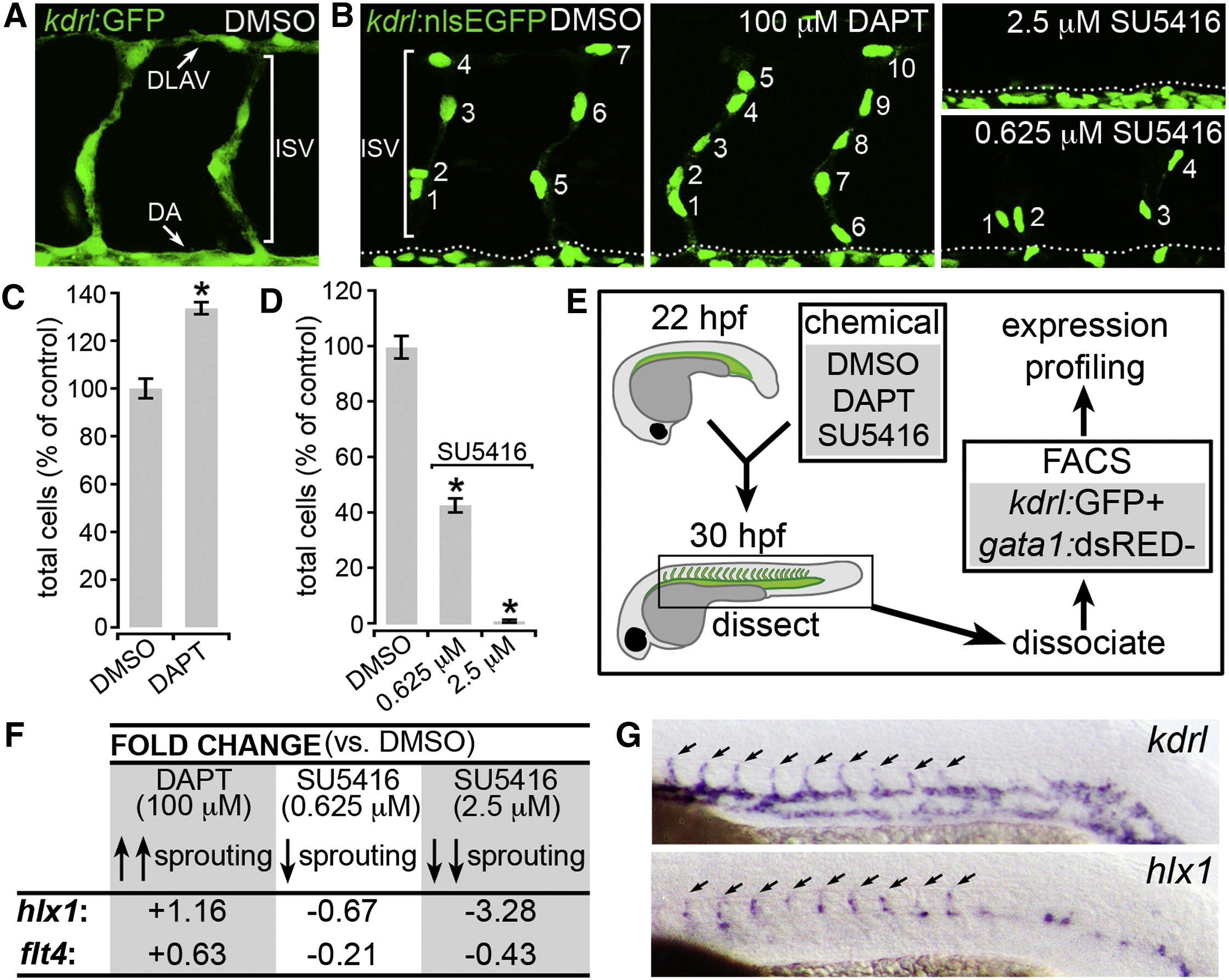
Fig. 1 hlx1 Expression Is Associated with Angiogenic Cell Behavior In Vivo(A and B) Lateral views of Tg(kdrl:GFP)s843 (A) or Tg(kdrl:nlsEGFP)zf109 (B) embryos at 30 hpf following incubation with either 0.4% DMSO, 100 μM DAPT, 2.5 μM SU5416, or 0.625 μM SU5416 from 22 hpf. Arrows in (A) indicate positions of the DA (dotted line in B denotes DA) and the forming DLAV, whereas white brackets in (A) and (B) indicate sprouting ISVs.(C and D) Quantification of ISV EC numbers at 30 hpf upon incubation of Tg(kdrl:nlsEGFP)zf109 embryos with either 100 μM DAPT (C) or the indicated concentration of SU5416 (D) (n = at least 21 embryos). A total of 100 μM DAPT augmented EC sprouting during ISV angiogenesis, whereas 2.5 and 0.625 μM SU5416 dose dependently disrupted EC-sprouting behavior.(E) Strategy for the identification of genes associated with EC-sprouting behavior in vivo. EC sprouting was pharmacologically manipulated prior to FACS-mediated isolation and transcriptome profiling of kdrl:GFP-positive ECs from dissected zebrafish trunks containing sprouting ISVs. Contaminating kdrl:GFP- and gata1:dsRed-double-positive erythrocytes were removed during FACS.(F) Fold change in EC hlx1 expression upon incubation with the indicated chemical versus DMSO controls. EC hlx1 expression was tightly correlated with the level of EC-sprouting behavior in vivo.(G) Whole-mount in situ hybridization analysis of the pan-endothelial marker kdrl or hlx1 at 24 hpf. hlx1 expression was enriched in sprouting ISVs (arrows).Error bars represent mean ± SEM. p < 0.05 versus control, Student’s t test.See also Figure S1.