
|
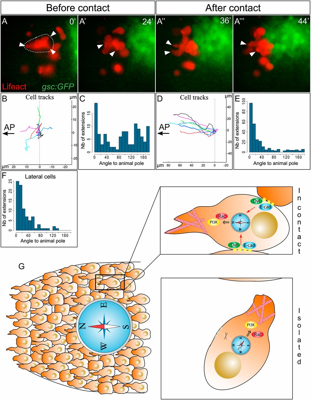
Fig. 4 Contact with the endogenous prechordal plate is required to orient migration. (A) Small groups of induced plate cells expressing Lifeact-mCherry were transplanted ahead of the endogenous prechordal plate in a Tg(gsc:GFP) embryo. Animal pole is to the left. Arrowheads point at cytoplasmic extensions. (B and D) Tracks of transplanted cells within isolated groups (B; Movie S8) or when the group has been contacted by the endogenous plate (D). Position of the cell at the onset of the movie (B) or at the time the group is contacted by the plate (D) has been set to position (0,0). (C and E) Orientation of the cytoplasmic extensions relative to the animal pole, when the group is isolated (C) or when it has been joined by the endogenous plate (E). (F) Orientation of cytoplasmic extensions relative to animal pole of cells located on lateral sides of the plate. If protrusion orientation was driven by contact inhibition alone, extensions should be perpendicular to the animal pole direction. Here they are oriented as in the rest of the plate (P > 0.05, n = 88 extensions). (G) Model of collective prechordal plate migration. All plate cells dynamically migrate toward the animal pole. Directionality is obtained by transmission of intrinsic information through cell–cell contacts. When isolated, cells show an autonomous ability to polarize, but they lose directionality.