
|
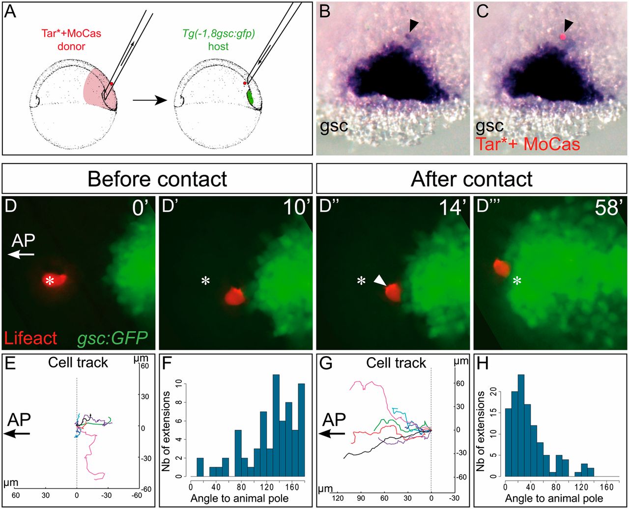
Fig. 2 Cell–cell contacts are required to orient migration. (A) Diagram of the experimental procedure. (B and C) A single induced plate cell expressing GFP (red in C) was transplanted ahead of the prechordal plate, at the onset of gastrulation. Although it is isolated, this cell keeps expressing goosecoid (B, black arrowhead). Here the embryo was fixed approximately 45 min after transplantation, just before the transplanted cell is contacted by the endogenous plate. Dorsal view is shown, with animal pole to the top. (D) A single induced plate cell expressing Lifeact-mCherry was transplanted ahead of the endogenous prechordal plate, in a Tg(gsc:GFP)embryo. Animal pole is to the left. Arrowhead points to an actin accumulation. Asterisk marks the position of the cell at the onset of the movie (Movie S7). (E and G) Tracks of transplanted cells while they remain isolated (E) or have been contacted by the endogenous plate (G). Position of the cell at the onset of the movie (E) or at the time it is contacted by the plate (G) has been set to position (0,0). (F and H) Orientation of the cytoplasmic extensions relative to the animal pole, when cells are isolated (F) or have been joined by the endogenous plate (H).