
|
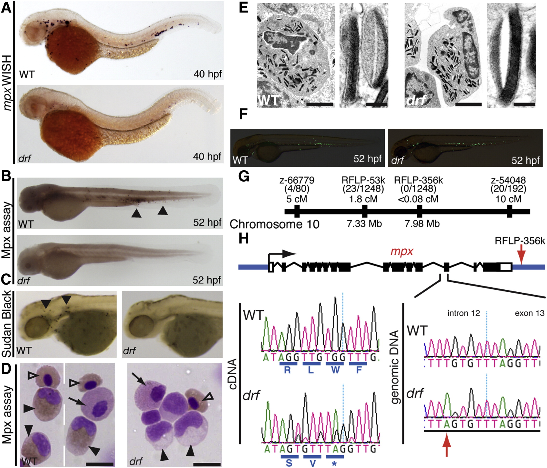
Fig. 2 Myeloperoxidase (mpx) Deficiency in the Zebrafish Mutant durif (drf)(A) mpx expression by whole-mount in situ hybridization (WISH) in WT and drf embryos.(B and C) Loss of cells carrying Mpx activity in drf demonstrated by loss of Mpx-dependent histochemical staining (B) and stable sudanophilia (C) (arrowheads indicate positive cells in WT).(D) Cytospun adult kidney hematopoietic cells, stained for Mpx by diaminobenzidine (DAB) histochemistry with May-Grunwald/Giemsa counterstain. WT but not drf neutrophils show Mpx staining (black triangles). Providing negative and positive internal staining controls, both genotypes have Mpx-negative eosinophils (black arrow) and weak erythrocyte staining due to the pseudoperoxidase Lepehne reaction (open triangles). Scale bar represents 10 μm.(E) Ultrastructure of adult WT and drf neutrophils, showing normal primary granules. Scale bars represent left, 2 μm; right, 100 nm.(F) drf-/- Tg(mpx:EGFP) hemizygotes have normal numbers of EGFP-expressing cells (mean ± SE: WT, 120 ± 6, n = 16; drf, 116 ± 5, n = 15).(G) Positional cloning placed drf on chromosome 10 within 0.08 cM of RFLP-356k located ~1.5 kb 3′ from the mpx gene.(H) Schematic of the mpx locus (black, exon; white, UTR; based on GenBank BC068379.1). Left sequencing chromatograms of complementary DNA shows the exon 11/12 junction: a 7 nt insertion in drf introduces a premature stop codon (*). Right sequencing chromatograms of genomic DNA show the drf mutation (red arrow), a T→A transversion 9 nt 52 of the intron 12/exon 13 junction.