
Fig. 5
- ID
- ZDB-IMAGE-121210-18
- Antibodies
- Publication
- Shin et al., 2012 - Zebrafish neurofibromatosis type 1 genes have redundant functions in tumorigenesis and embryonic development
- All Figures
- Figures for Shin et al., 2012
|
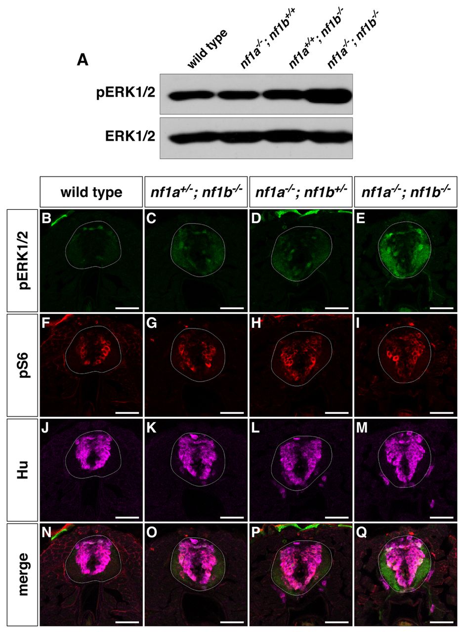
Fig. 5 nf1a/nf1b mutants exhibit upregulation of pERK1/2. (A) Western blot analysis for pERK1/2 in protein lysates prepared from wild-type, nf1a-/-; nf1b+/+, nf1a+/+; nf1b-/- and nf1a-/-; nf1b-/- larvae (100 μg) reveals increased pERK1/2 levels in nf1a-/-; nf1b-/- larvae as compared with wild-type, nf1a-/-; nf1b+/+ and nf1a+/+; nf1b-/- larvae at 3 dpf. Equal loading was confirmed by stripping the membrane and reprobing for total ERK1/2. (B-Q) Transverse spinal cord sections of wild-type (B,F,J,N), nf1a+/-; nf1b-/- (C,G,K,O), nf1a-/-; nf1b+/- (D,H,L,P) and nf1a-/-; nf1b-/- (E,I,M,Q) larvae labeled with anti-pERK1/2 antibody (B–E, green), anti-pS6 antibody (F-I, red), or anti-HuC/D antibody (J-M, magenta) demonstrate marked upregulation of pERK1/2 in nf1a-/-; nf1b-/-larvae and intermediate levels of pERK1/2 in larvae harboring a single functioning nf1 allele as compared with wild-type larvae at 4 dpf (n=5 each for wild-type and mutant larvae). Scale bars: 40 μm.