
|
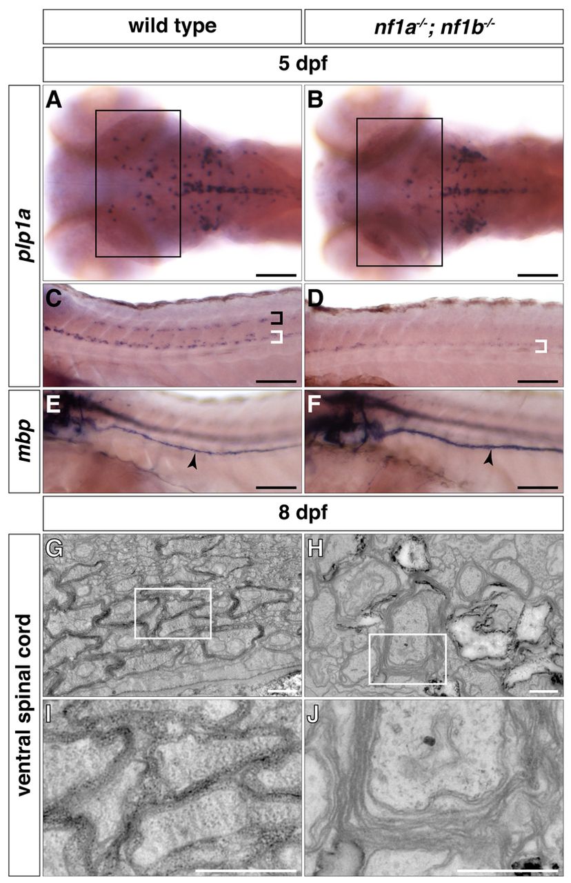
Fig. 4 nf1a-/-; nf1b-/- larvae exhibit myelination defects. (A–D) plp1a expression is decreased in glial cells of the midbrain and hindbrain regions (A,B, boxes) as well as the dorsal (black bracket) and ventral (white brackets) spinal cord of nf1a-/-; nf1b-/- larvae (B,D) as compared to wild-type larvae (A,C) by whole-mount in situ hybridization at 5 dpf. (E,F) mbp expression is elevated in Schwann cells of the PLLn (arrowheads) of nf1a-/-; nf1b-/- larvae (F) as compared to wild-type larvae (E) by whole-mount in situ hybridization at 5 dpf. (G-J) Transverse sections through the ventral spinal cord nf1a-/-; nf1b-/-larvae (H, boxed region magnified in J) demonstrate defects in formation of compact myelin sheaths as compared with wild-type larvae (G, boxed region magnified in I) at 8 dpf. Scale bars: 100 μm (A-F) and 0.5 μm (G-J).