
Fig. 7
- ID
- ZDB-IMAGE-121205-9
- Publication
- Blaker-Lee et al., 2012 - Zebrafish homologs of 16p11.2, a genomic region associated with brain disorders, are active during brain development, and include two deletion dosage sensor genes
- All Figures
- Figures for Blaker-Lee et al., 2012
|
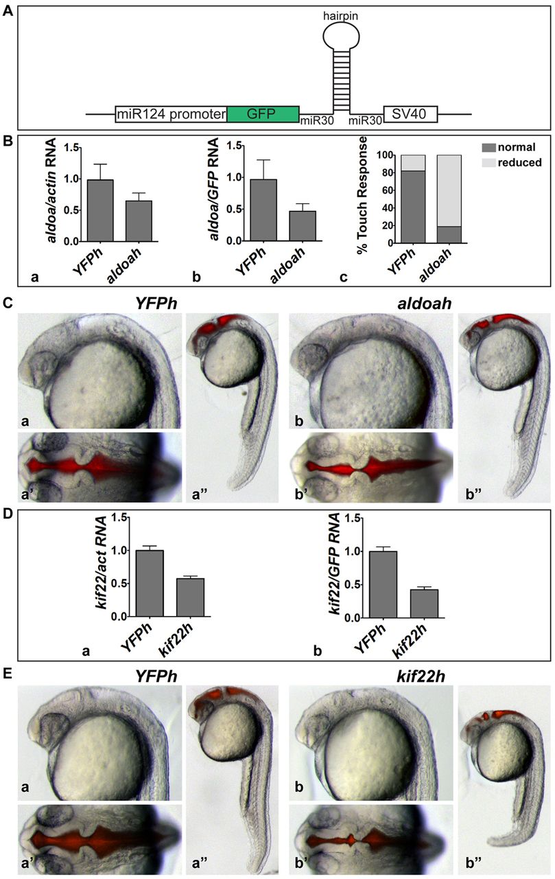
Fig. 7 aldoaa and kif22 function are required in the brain. (A) shRNA expression strategy. shRNAs were expressed from a miR30 backbone, under the CNS-specific miR124 promoter. Transient transgenesis was induced using I-SceI meganuclease. (B) Relative expression of aldoaa mRNA after inhibition by shRNA. Expression was quantified by qPCR in 24 hpf embryos injected with an aldoaa hairpin (aldoah), shown relative to embryos injected with a control hairpin (YFPh). Data was normalized to either (Ba) actin or (Bb) GFP expression. (Bc) Touch response in 24 hpf aldoah embryos versus control YFPh embryos (81% abnormal, n=26). (C) Phenotype of shRNA-injected embryos. (Ca-Ca′′) 24 hpf control YFPh embryos (16% abnormal, n=51, in two independent experiments). (Cb-Cb′′) 24 hpf aldoah embryos (89% abnormal, n=53, in two independent experiments). (Ca,Cb) Lateral view of the head; (Ca′,Cb′) dorsal view of the head after brain ventricle injection; (Ca′′,Cb′′) lateral view of whole embryo. (D) Relative expression of kif22 mRNA after inhibition by shRNA. kif22 mRNA relative expression was quantified by qPCR in 24 hpf embryos injected with a kif22 hairpin (kif22h) relative to those injected with the YFPh control hairpin. Data was normalized to (Da) actin or (Db) GFP expression. (E) Phenotype of shRNA-injected embryos. (Ea-Ea′′) 24 hpf YFPh embryos (8% abnormal, n=27, in two independent experiments). (Eb-Eb′′) 24 hpf kif22h embryos (81% abnormal, n=26, in two independent experiments). (Ea,Eb) Lateral view of the head; (Ea′,Eb′) dorsal view of the head after brain ventricle injection; (Ea′′,Eb′′) lateral view of whole embryo.