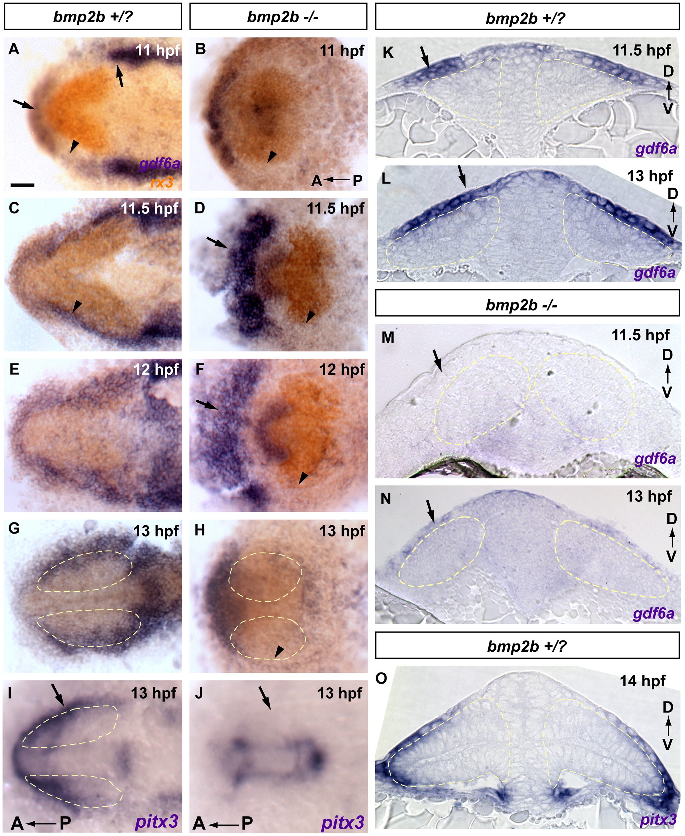
Fig. 6 Dorsal initiation signal arises from non-neural ectoderm at 11–12 hpf. Extraocular gdf6a is expressed in the correct time and place to initiate dorsal fate within the retina, and expression is absent in bmp2b mutants, lacking dorsal fate. These results suggest that lateral head ectoderm is the tissue responsible for initiating dorsal fate within the retina. (A–H) gdf6a expression in blue, rx3 expression in orange. (A, C and K) gdf6a in +/? siblings is expressed in the non-neural head ectoderm, and is initially strongest anterior and posterior to the optic vesicle (arrows in A, arrowhead in A marks weaker expression lateral to the optic vesicle). At 11.5 hpf gdf6a expression has intensified covering the lateral portion of the optic vesicle (C, arrowhead; K transverse section, arrow). (E, G and L) gdf6a later expands to cover the majority of the dorsal leaflet of the optic vesicle (G; L transverse section, arrow). (K and L) Extraocular gdf6a at 11.5–13 hpf is directly adjacent to optic vesicle tbx5a, initiated at 12 hpf (Compare K and L to Fig. 1A0–A3). (B, D, F, H, M and N) In bmp2b mutants the lateral extraocular domain of gdf6a is not detectable or greatly downregulated (arrowheads in B, D, F and H, arrows in M and N). Expression of gdf6a anterior to the optic vesicle is upregulated in mutants (arrows in D and F). (I, J and O) The extraocular lateral domain of the non-neural ectoderm and lens placode marker pitx3 is also absent in bmp2b mutants (arrows in I and J), indicating that bmp2b mutants do not form the non-neural ectodermal tissue necessary for gdf6a expression and subsequent initiation of dorsal fate. (A–J) whole mount dorsal view, anterior to the left. (K–O) transverse sections. Scale bars=50 μM. Dashed yellow lines outline optic vesicles.
Reprinted from Developmental Biology, 371(1), Kruse-Bend, R., Rosenthal, J., Quist, T.S., Veien, E.S., Fuhrmann, S., Dorsky, R.I., and Chien, C.B., Extraocular ectoderm triggers dorsal retinal fate during optic vesicle evagination in zebrafish, 57-65, Copyright (2012) with permission from Elsevier. Full text @ Dev. Biol.

