
|
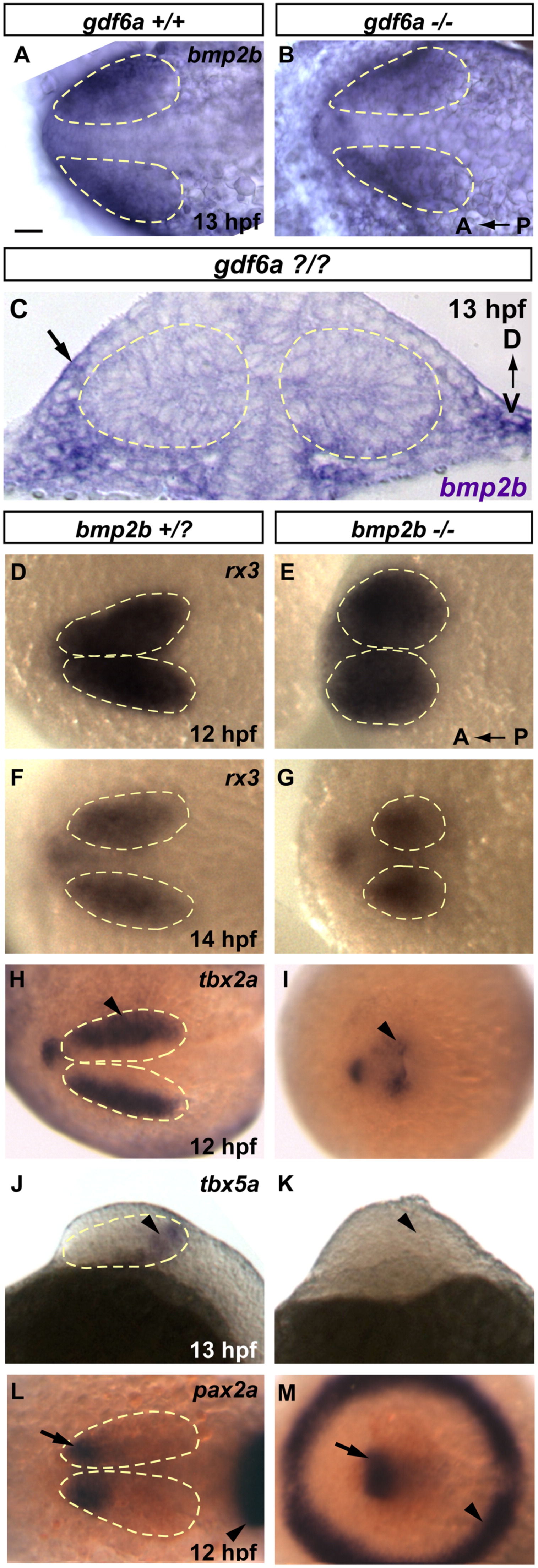
Fig. 5 bmp2b is necessary for dorsal retina initiation. Despite severe morphological defects, bmp2b mutants develop rx3 expressing eye fields. They fail, however to initiate expression of several dorsal markers. (A–C) bmp2b is expressed in the extraocular ectoderm in 13 hpf gdf6a siblings (A) and this expression domain is unchanged in gdf6a mutants (B). (D–G) rx3 expression in siblings and bmp2b mutants at 12 and 14 hpf. (H and I) The earliest dorsal fate marker, tbx2a, is expressed in siblings beginning at 11 hpf (12 hpf shown, arrowhead). tbx2a is greatly downregulated in bmp2b mutants (H, arrowhead). (J and K) tbx5a expression is initiated at 12 hpf in siblings (13 hpf shown, arrowhead in I), but never turns on in bmp2b mutants (arrowhead in K where staining is absent). (L and M) In 12 hpf siblings, pax2a marks the prospective ventral retinal domain (arrow, L), which is also present in bmp2b mutants at this time point (arrow, M), despite severe dorsalization, which leads to a circular midbrain–hindbrain boundary (arrowheads; compare M with L). A and B, D–I, L and M are dorsal views, anterior to the left. J and K are lateral views, anterior to the left; C is a transverse section. Scale bars=50 μM. Dashed yellow lines outline optic vesicles where boundaries are visible.
Reprinted from Developmental Biology, 371(1), Kruse-Bend, R., Rosenthal, J., Quist, T.S., Veien, E.S., Fuhrmann, S., Dorsky, R.I., and Chien, C.B., Extraocular ectoderm triggers dorsal retinal fate during optic vesicle evagination in zebrafish, 57-65, Copyright (2012) with permission from Elsevier. Full text @ Dev. Biol.