
|
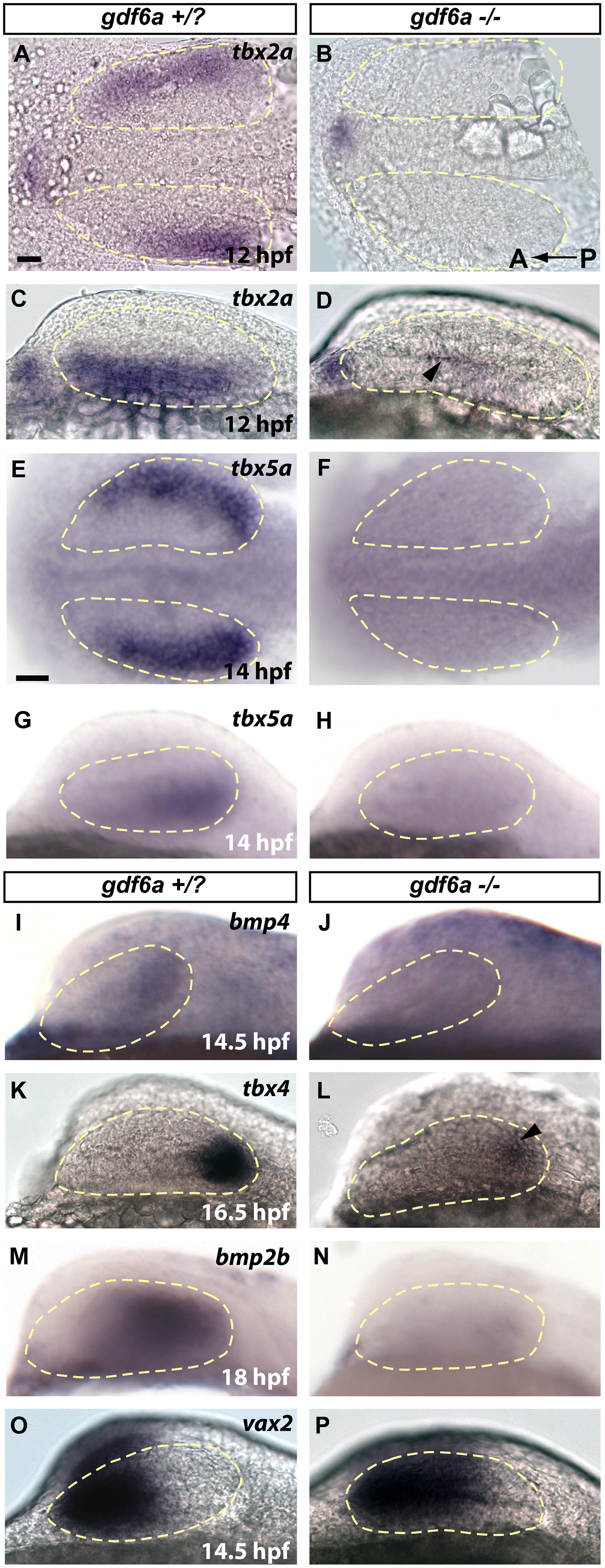
Fig. 4 gdf6a is necessary for establishing dorsal retinal fate. In gdf6a mutants, dorsal retinal patterning genes either fail to express, or have greatly downregulated expression. Ventral genes are turned on normally, but expand shortly after to fill almost the entire retinal field. Expression of dorsally or ventrally expressed genes is shown at stages shortly after they are first detectable in wild type. (A–D) Robust 12 hpf tbx2a in gdf6a siblings is only faintly detectable in gdf6a mutants (arrowhead in D). (E–H) Expression of tbx5a begins at 12 hpf (14 hpf shown in figure) in sibling embryos, but is never seen in gdf6a mutants (F and H). (I–N) Similarly, bmp4 and bmp2b are completely lost in gdf6a mutants, while tbx4 expression is initiated at very low levels (arrowhead in L). (O and P) Expression of vax2 is only slightly expanded at 14.5 hpf, and continues to expand to fill most of the retina by 24 hpf (See supplementary Fig. 3R) Anterior to the left; A, B, E and F are dorsal views; C, D, G–R are lateral views. Scale bars=50 μM. Dashed yellow lines outline optic vesicles.
Reprinted from Developmental Biology, 371(1), Kruse-Bend, R., Rosenthal, J., Quist, T.S., Veien, E.S., Fuhrmann, S., Dorsky, R.I., and Chien, C.B., Extraocular ectoderm triggers dorsal retinal fate during optic vesicle evagination in zebrafish, 57-65, Copyright (2012) with permission from Elsevier. Full text @ Dev. Biol.