
|
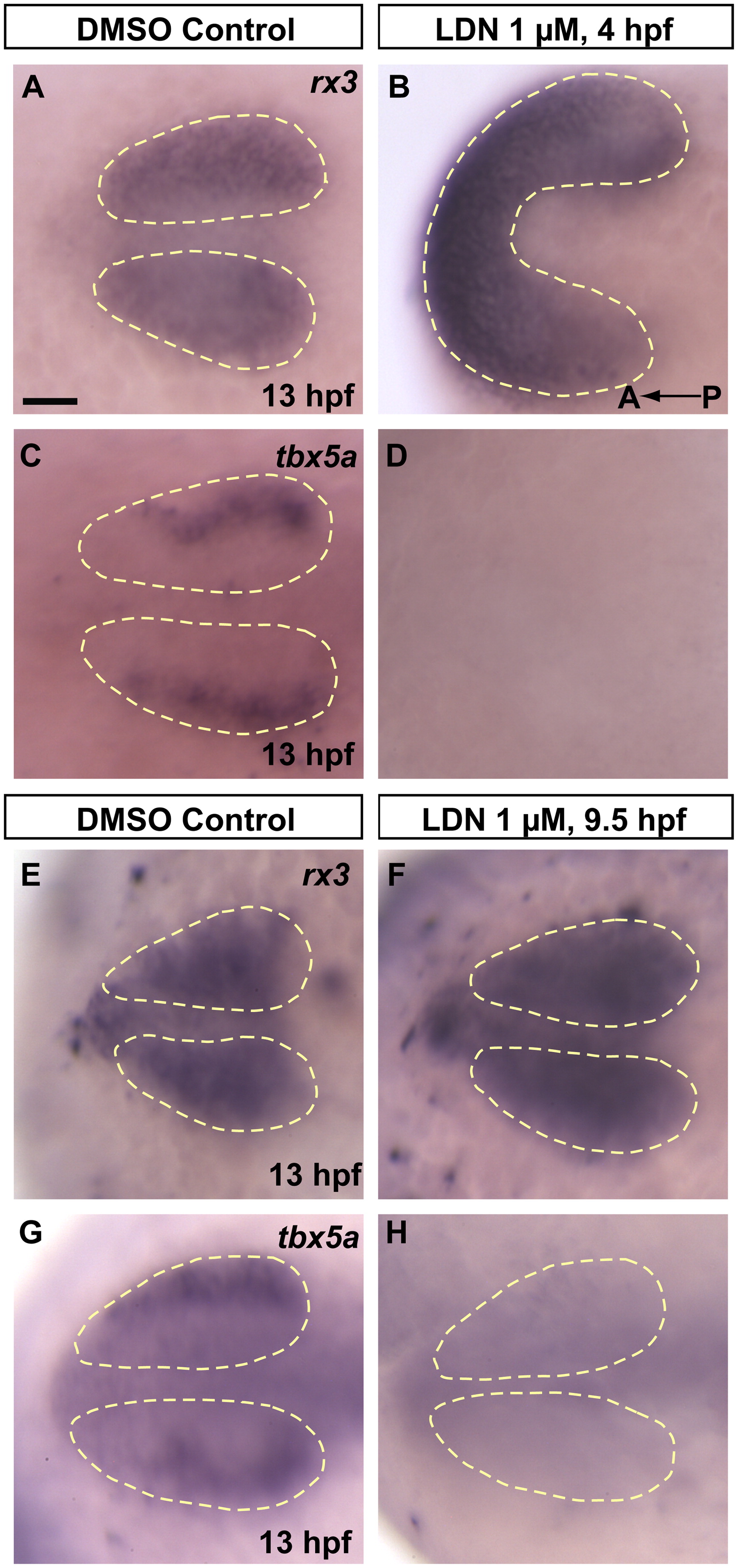
Fig. 3 bmp signals necessary for dorsal fate initiation are acting between 9.5 hpf and 13 hpf. Pharmacological inhibition of bmp signaling using the small molecule inhibitor LDN 193189 prevents initiation of dorsal fate in the retina. (A and B) rx3 expression following 4–13 hpf continuous treatment with 1% DMSO in E2/GN (control) or 1 μM LDN 193189 in 1% DMSO E2/GN shows that treatment with LDN 193189 leads to severely dorsalized embryos, yet these embryos still form an eye field. (C and D) Contrary to DMSO controls, eye fields in LDN treated embryos fail to initiate expression of the prospective dorsal marker tbx5a. (E and F) Embryos treated from 9.5–13 hpf with 1% DMSO in E2/GN (control) or 1 μM LDN 193189 in 1% DMSO E2/GN both show normal rx3 expression, indicating normal eye field formation. (G and H) However, contrary to controls, optic vesicles in treated embryos fail to initiate expression of the prospective dorsal marker tbx5a, suggesting that dorsal fate is never initiated if bmp signaling is inhibited after 9.5 hpf. All images are dorsal views, anterior to the left. Scale bar=50 μM. Dashed yellow lines outline optic vesicle domains.
Reprinted from Developmental Biology, 371(1), Kruse-Bend, R., Rosenthal, J., Quist, T.S., Veien, E.S., Fuhrmann, S., Dorsky, R.I., and Chien, C.B., Extraocular ectoderm triggers dorsal retinal fate during optic vesicle evagination in zebrafish, 57-65, Copyright (2012) with permission from Elsevier. Full text @ Dev. Biol.