
|
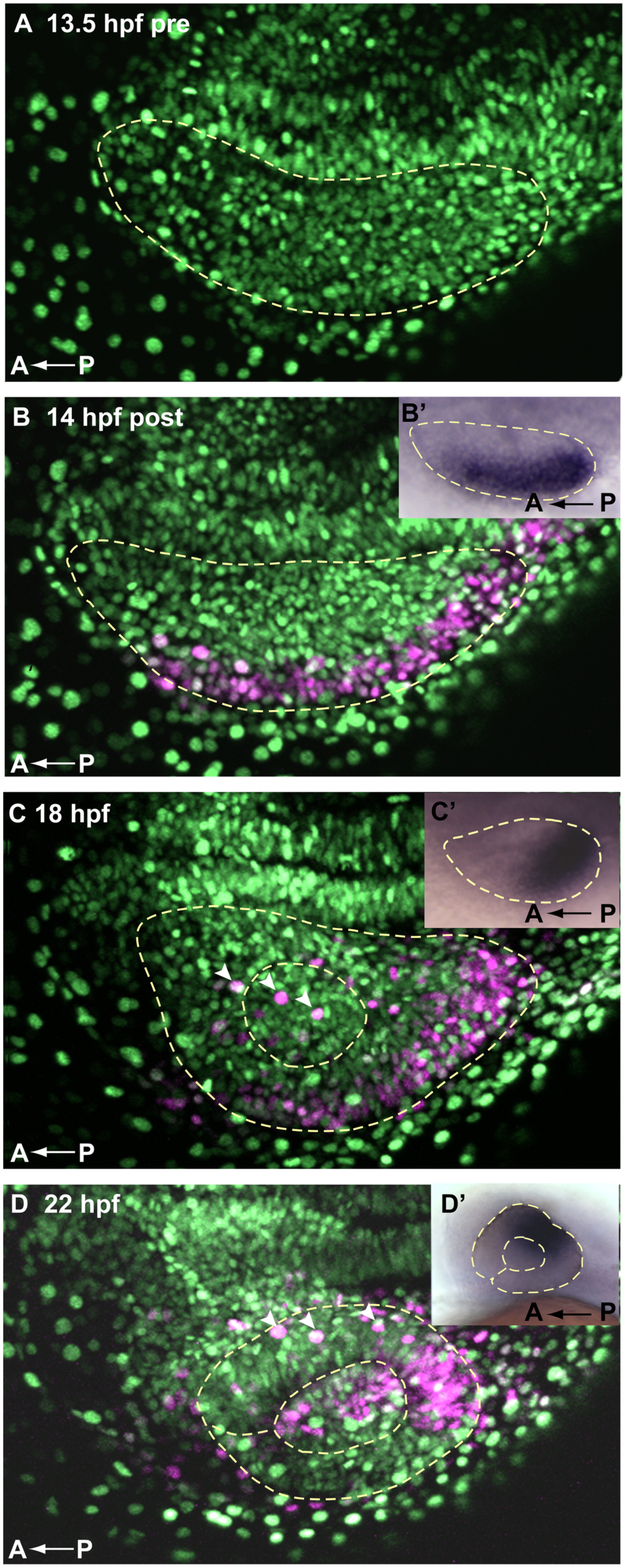
Fig. 2 Cells of the lateral optic vesicle give rise to prospective dorsal and central retina. A Kaede fate map of 14 hpf lateral retina shows that cell movements generally recapitulate movements of tbx5a expression, indicating that dorsal fate initiation could be a discrete early event. (A–D) Dorsal view, maximum intensity projections of single optic vesicles, oriented as shown in the schematic (Fig. 1 A0–D0′) with the midline toward the top. Dashed yellow lines outline optic vesicles (A–D′), lenses (C–D′), and choroid fissure (D and D′). (A) Pre-activation, ubiquitous NLS green Kaede, 13.5 hpf. (B) 14 hpf, immediately after photoactivation, cells of lateral optic vesicle express photoactivated Kaede (magenta). (B′) 14 hpf whole mount tbx5a expression, dorsal view (C) 18 hpf, photoactivated Kaede cells from lateral optic vesicle have moved posteriorly. (C′) 18 hpf whole mount tbx5a expression, dorsal view (D) 22 hpf, photoactivated magenta cells reside primarily in the dorsal and central retinal domains, as well as in the lens. (D′) 24 hpf whole mount tbx5a expression, lateral view. Movements of photocleaved Kaede 14 hpf lateral retinal cells are generally consistent with the pattern of movement of tbx5a—compare magenta domains in A–D to A′–D′ insets and to Fig. 1 sections. White arrowheads in C and D mark extraocular ectodermal cells overlying the retina and lens.
Reprinted from Developmental Biology, 371(1), Kruse-Bend, R., Rosenthal, J., Quist, T.S., Veien, E.S., Fuhrmann, S., Dorsky, R.I., and Chien, C.B., Extraocular ectoderm triggers dorsal retinal fate during optic vesicle evagination in zebrafish, 57-65, Copyright (2012) with permission from Elsevier. Full text @ Dev. Biol.