
|
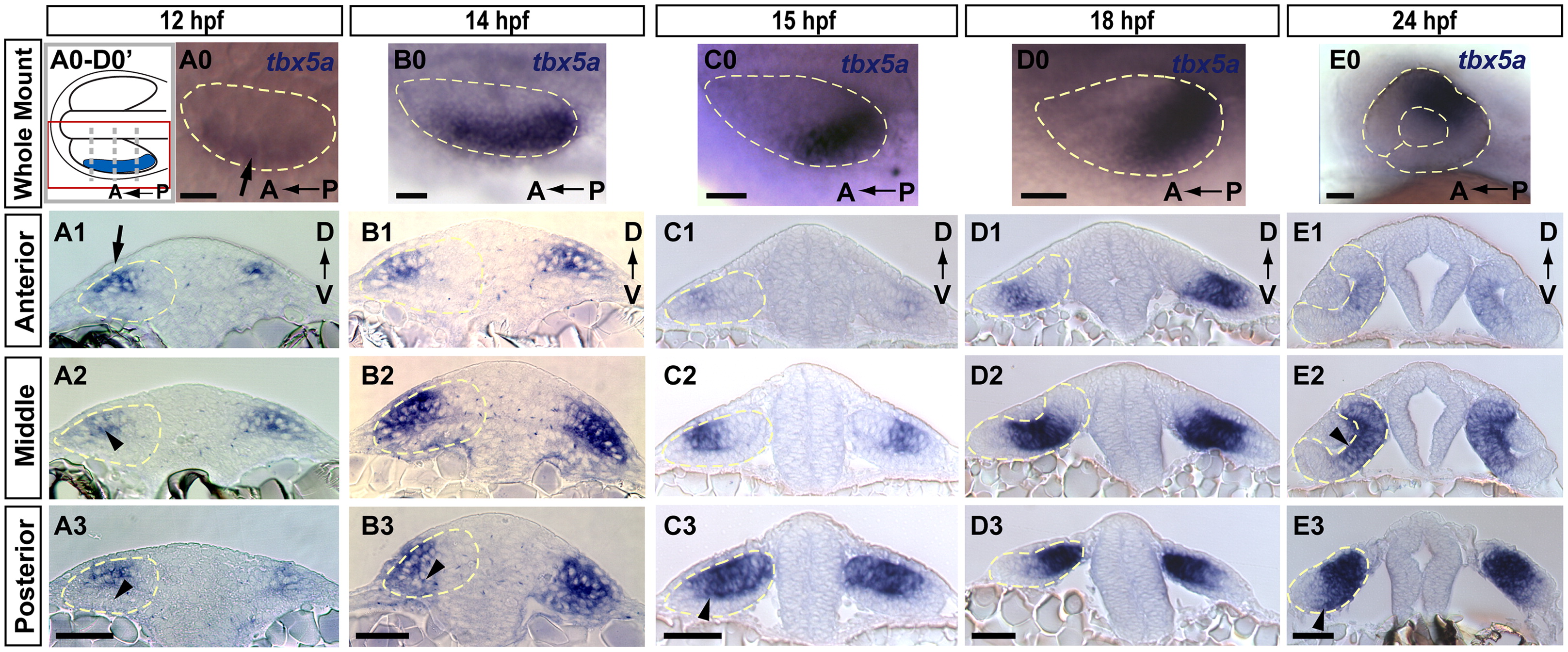
Fig. 1 Expression domain of prospective dorsal marker tbx5a is broad and dynamic. Expression of tbx5a in the developing retina at (A) 12, (B) 14, (C) 15, (D) 18 and (E) 24 hpf (blue). (0 whole mount; 1–3, transverse sections). (A0–D0 dorsal views; E0, lateral view). (A0–D0′) Schematic diagram of orientation for A0–D0 whole-mount dorsal view images. Each image shows a single optic vesicle (red box in schematic), with the midline oriented toward the top. Dashed gray lines show approximate location of anterior, middle, and posterior sections in 1–3. (A) tbx5a mRNA is expressed in dorsolateral optic vesicle (arrow in A0, arrowhead in A2) at 12 hpf, and is absent from the ventral leaflet (arrowhead in A3). Extraocular ectoderm is directly overlying this portion of the optic vesicle (arrow in A1). (B) At 14 hpf expression extends to the ventral leaflet in posterior sections (arrowhead in B3). (C and D) tbx5a domain moves posteriorly from 15–18 hpf and resides primarily in the dorsal (prospective retinal) leaflet (arrowhead in C3). (E) tbx5a expression still covers much of the eye at 24 hpf, including prospective dorsal pole, central retinal domain medial to the lens (arrowhead in E2), and extending to the ventral domain (arrowhead in E3). Scale bars=50 μM. Dashed yellow lines outline optic vesicles in all images, also lens and choroid fissure in E0.
Reprinted from Developmental Biology, 371(1), Kruse-Bend, R., Rosenthal, J., Quist, T.S., Veien, E.S., Fuhrmann, S., Dorsky, R.I., and Chien, C.B., Extraocular ectoderm triggers dorsal retinal fate during optic vesicle evagination in zebrafish, 57-65, Copyright (2012) with permission from Elsevier. Full text @ Dev. Biol.