
|
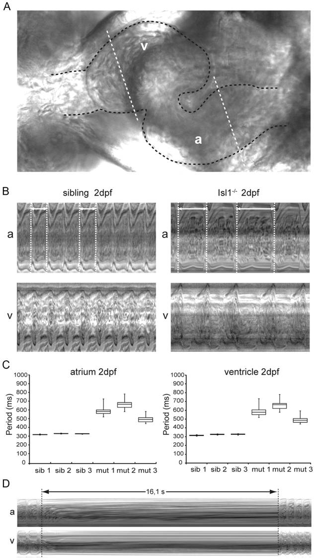
Fig. 1 Characterization of the embryonic Isl1-/- cardiac phenotype in vivo.
(A) Zebrafish embryonic heart at 2 dpf. The embryonic heart is highlighted in black dotted contour; white dotted lines through the atrium (A) and the ventricle (V) are placed at kymograph positions. (B) Atrial (A) and ventricular (V) kymographs from 2 dpf embryonic hearts spanning approximately 2.8 s. Note the much longer period of the Isl-/- heart when compared to the sibling and the irregularity of the period (double arrow and white dotted vertical lines). Movies are available as Movies S1 and S2, respectively. (C) Box-whisker plots representation of 20 successive heartbeats of 2 dpf Isl1-/- and sibling embryos. (D) Kymograph recorded at 3 dpf covering a period of about 16 s of absent heart contractions. For all panels a: atrium; v: ventricle.