
|
Fig. S2
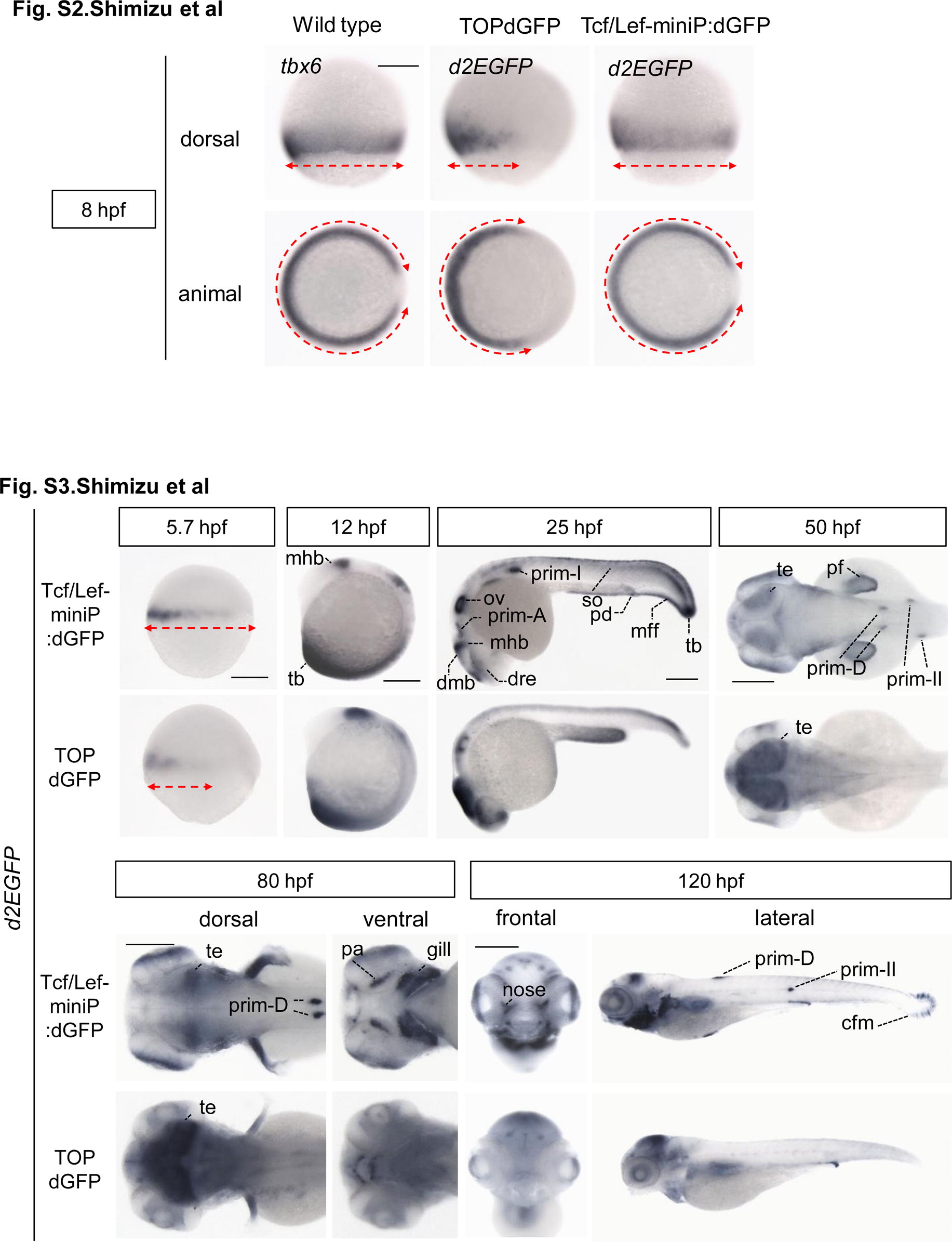
The expression pattern of the Tcf/Lef-miniP:dGFP reporter at 8 hpf, but not that of the TOPdGFP reporter, was completely consistent with that of tbx6 mRNA. Whole-mount in situ hybridization staining for tbx6 in wild-type zebrafish embryos and for d2EGFP mRNA in Tcf/Lef-miniP:dGFP and TOPdGFP transgenic zebrafish embryos at 8 hpf. The expression domains of tbx6 and d2EGFP mRNAs are indicated with red broken lines. Tcf/Lef-miniP:dGFP reporter activity and tbx6 mRNA were detected in both the ventral and dorsal lateral regions. In contrast, TOPdGFP reporter activity was observed only in the ventral region but not in the dorsal lateral region, indicating that the expression pattern was not completely consistent with that of tbx6 mRNA. and Figure S3. Comparison of the expression pattern of d2EGFP mRNA in Tcf/Lef-miniP:dGFP and TOPdGFP transgenic zebrafish embryos. Left-side (panels for 5.7, 12, and 25 hpf, and right panel for 120 hpf), dorsal (panels for 50 hpf and left panels for 80 hpf), ventral (right panels for 80 hpf), and frontal (left panels for 120 hpf) views of Line-2 or TOPdGFP-transgenic zebrafish embryos, with the ventral side to the left (panels for 5.7 and 12 hpf), the anterior side to the left (panels for 25, 50, and 80 hpf, and right panels for 120 hpf), or the dorsal side to the top (left panels for 120 hpf). Scale bar: 200 μm. The expression domains of d2EGFP mRNA at 5.7 hpf are indicated with red broken lines. The expression domains of tbx6 and d2EGFP mRNAs are indicated with red broken lines. At 5.7 hpf, the d2EGFP mRNA-expressing domain in TOPdGFP transgenic fish was narrower than that in Line-2. d2EGFP mRNA expression was detected in the median and pectoral fin fold (mff and pff), caudal fin mesenchymal cell (cfm), prim-A, prim-II, prim-D, developing pharyngeal arch (pa), gill, and nose in Line-2 embryos, but not in TOPdGFP fish embryos.
Reprinted from Developmental Biology, 370(1), Shimizu, N., Kawakami, K., and Ishitani, T., Visualization and exploration of Tcf/Lef function using a highly responsive Wnt/beta-catenin signaling-reporter transgenic zebrafish, 71-85, Copyright (2012) with permission from Elsevier. Full text @ Dev. Biol.