
Image
|
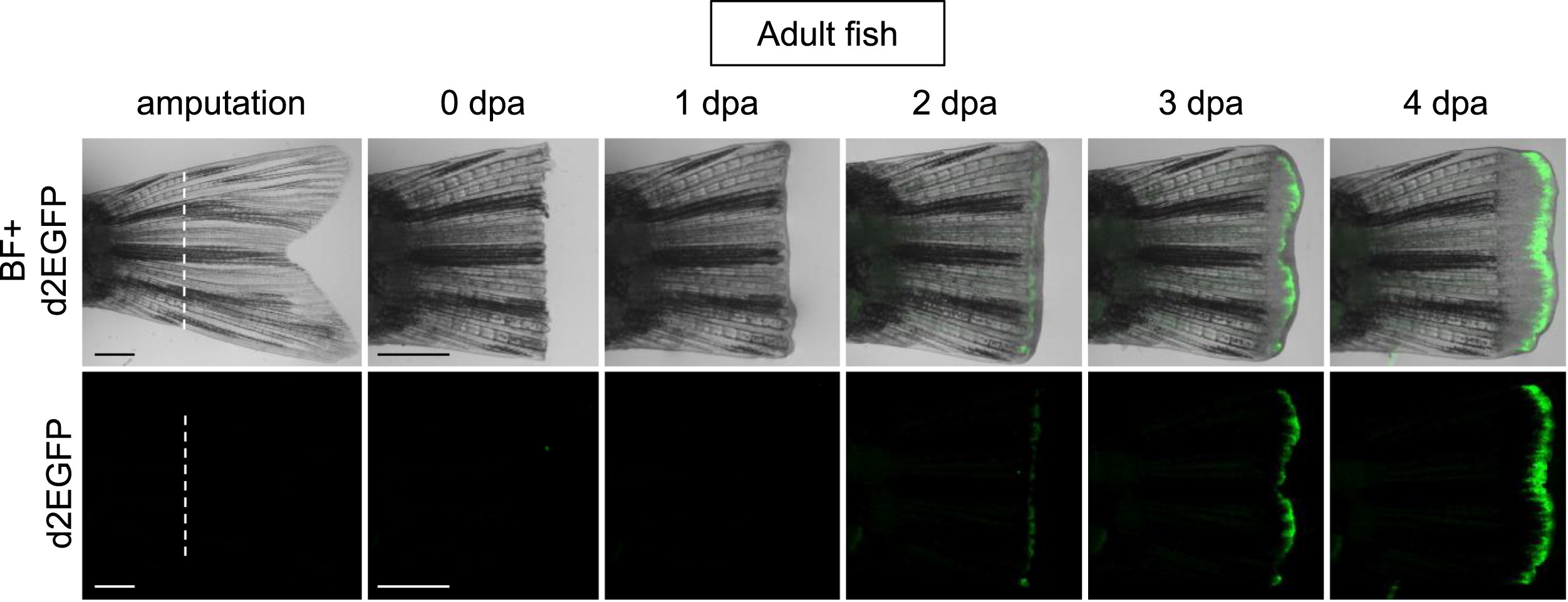
Figure Caption
Fig. 5
Tcf/Lef-miniP:dGFP reporter was activated in the amputated caudal fin regenerative outgrowths of adult transgenic fish. The caudal fin was amputated at the white line indicated, as shown for 4 dpa (0–4 dpa). d2EGFP-expressing cells were visualized by fluorescence microscopy (lower panels). The fluorescence images were merged with bright-field (BF) images (upper panels). The Tcf/Lef-miniP:dGFP reporter was upregulated in the amputated fin during regeneration. Scale bar, 200 μm.
Acknowledgments
This image is the copyrighted work of the attributed author or publisher, and
ZFIN has permission only to display this image to its users.
Additional permissions should be obtained from the applicable author or publisher of the image.
Reprinted from Developmental Biology, 370(1), Shimizu, N., Kawakami, K., and Ishitani, T., Visualization and exploration of Tcf/Lef function using a highly responsive Wnt/beta-catenin signaling-reporter transgenic zebrafish, 71-85, Copyright (2012) with permission from Elsevier. Full text @ Dev. Biol.