
|
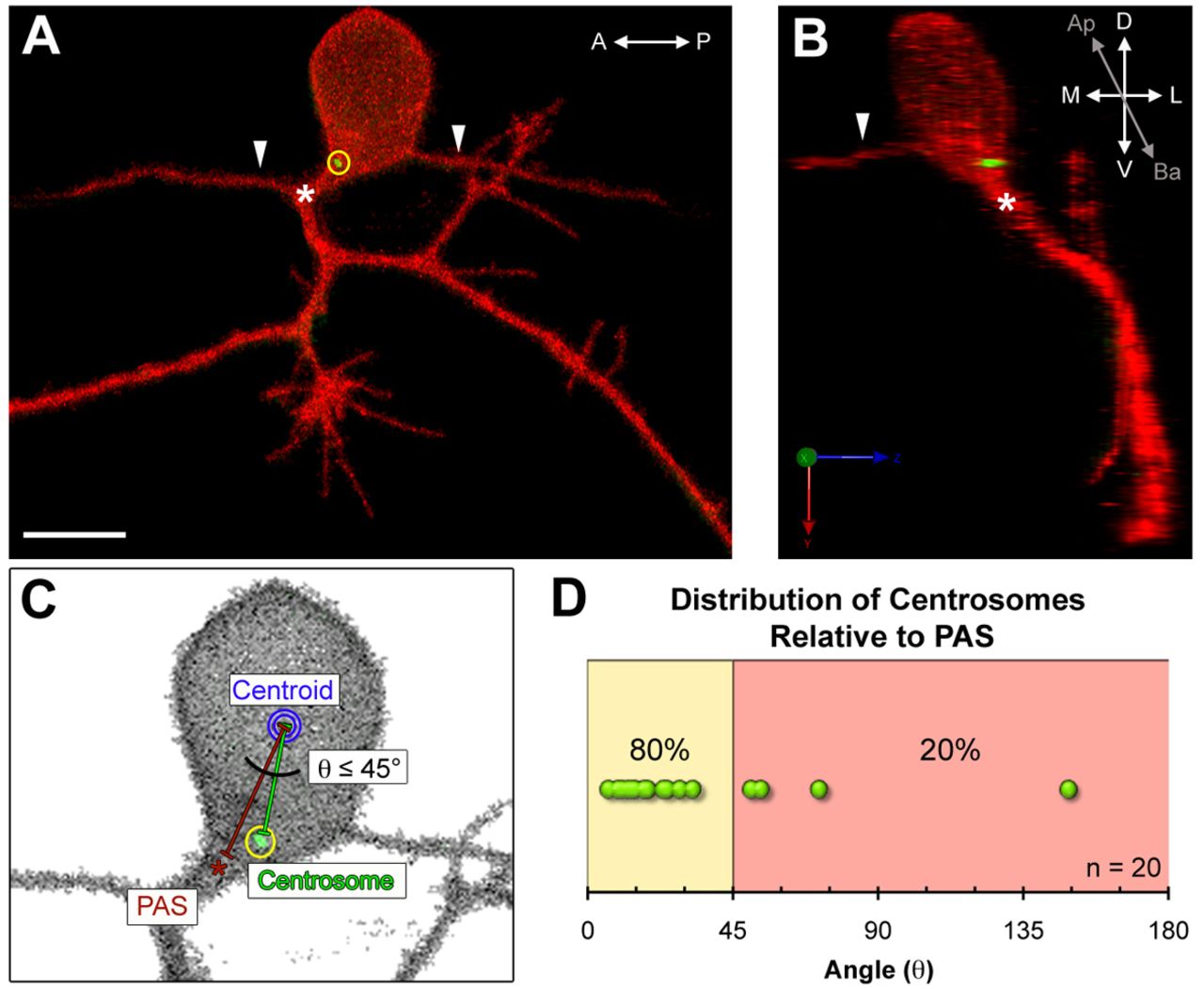
Fig. 1 The centrosome is positioned near the peripheral axon initiation site in mature Rohon-Beard neurons. (A,B) Images of a mature Rohon-Beard (RB) neuron labeled by transient expression of TagRFP-CAAX in a wild-type zebrafish embryo expressing GFP-Xcentrin mRNA. (A) z-projection showing central axons extended along the anterior-posterior (A-P) axis (arrowheads) and a branched peripheral axon orthogonal to the A-P axis. The centrosome (yellow circle) is localized near the peripheral axon initiation site (PAS, asterisk). Scale bar: 10 μm. (B) Three-dimensional rendering of the neuron in A (yz view) showing the centrosome at the basal-lateral (Ba-L) cell body surface, near the PAS. (C) Schematic showing how centrosome position (yellow circle) was measured relative to the PAS and centroid (blue circle). (D) Distribution of angle (θ) measurements defined in C. The line between the yellow and red zones indicates 45° angle cut-off.