
|
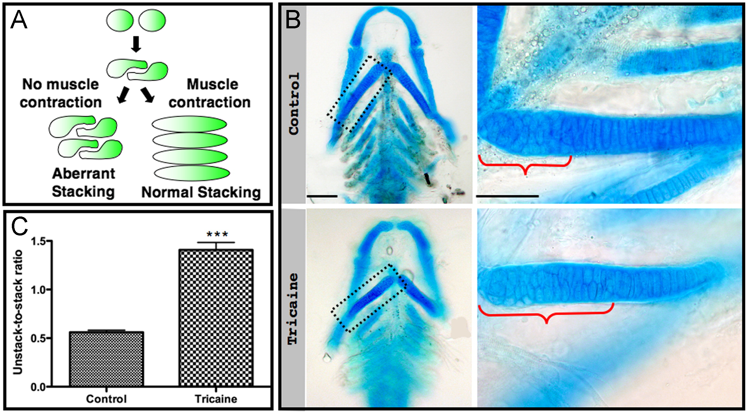
Fig. 4 Short-term paralysis results in aberrant stacking. (A) A model illustrating the involvement of muscle contraction in chondrocyte intercalation (modified from Li and Dudley, 2009). (B) Flat-mounted Alcian blue stained control (upper panel) and tricaine-paralyzed (lower panel) 120 hpf zebrafish. Magnifications of the boxed areas are shown on the right. Brackets indicate the length of the area occupied by unstacked chondrocytes. Scale bars are 100 μm on the left and 50 μm on the right. (C) Quantification of the ratio of the lengths of areas of unstacked to stacked chondrocytes in four controls and four paralyzed zebrafish (p<0.0001).
Reprinted from Developmental Biology, 370(1), Shwartz, Y., Farkas, Z., Stern, T., Aszódi, A., and Zelzer, E., Muscle contraction controls skeletal morphogenesis through regulation of chondrocyte convergent extension, 154-163, Copyright (2012) with permission from Elsevier. Full text @ Dev. Biol.