
|
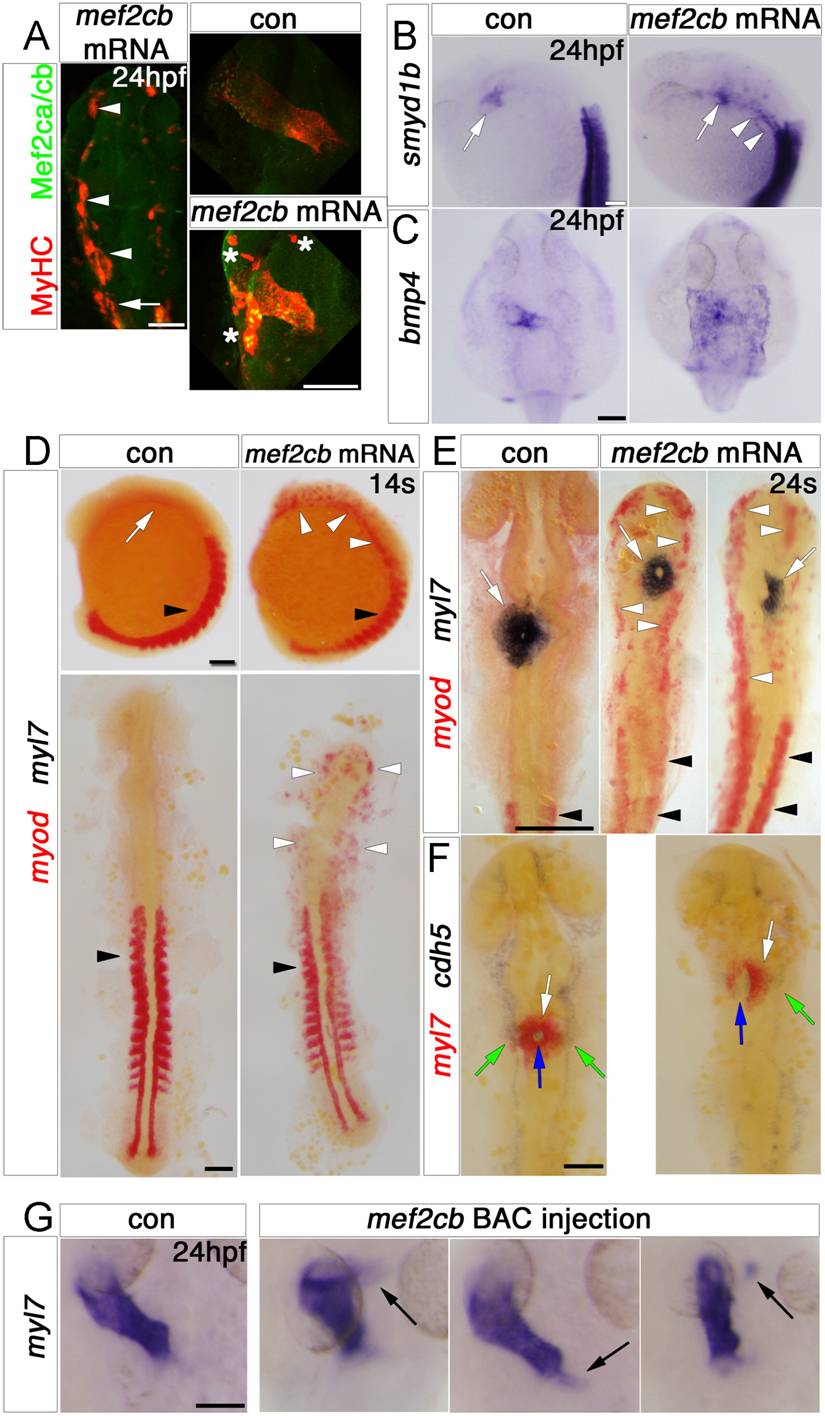
Fig. 6 Mef2cb overexpression induces skeletal muscle at the expense of myocardial and endothelial cells. In situ mRNA hybridisation (or immunodetection, A) for indicated genes, shown as wholemounts in dorsal (G and C) or lateral (B and D, top panel) views, or as flatmounts in dorsal view (A and D, bottom panel, E and F). A. Injection of mef2cb RNA results in many ectopic muscle cells expressing strong Mef2ca/cb and MyHC at 24 hpf (white arrowheads) anterior to the first somite (white arrow). In the heart, MyHC and Mef2ca/cb are mosaically stronger than in control embryos. Asterisks=ectopic muscle. B.Smyd1b is upregulated in CMs (white arrow), and in ectopic muscle in the head region (white arrowheads). C. Expression of bmp4 is upregulated in much of a sheet of cardiogenic cells but not elsewhere in the embryo. D. In 14 ss control embryos, myod mRNA is expressed in somites (black arrowheads) and myl7 is expressed weakly in CMs (white arrow). Embryos injected with mef2cb RNA express no ectopic myl7, but have ectopic myod mRNA in the head region (white arrowheads). E. By 24 ss, high levels of ectopic myod correlated with reduction or lack of myl7-expressing CMs (right panel) and defective brain development. F. At 24 ss, mef2cb RNA-injected embryos that had fewer myl7-expressing CMs (white arrows) also had less cdh5 expression in vascular endothelium (green arrows) and disorganised endocardium (blue arrows). G. Compared to wild type control (leftmost panel), three examples of embryos injected with mef2cb BAC DNA show ectopic myl7 mRNA either contiguous with (left panels) or detached from (right panel) the arterial (flanking panels) or venous (middle panel) poles. Scale=100 μm. (For interpretation of the references to color in this figure legend, the reader is referred to the web version of this article.)
Reprinted from Developmental Biology, 369(2), Hinits, Y., Pan, L., Walker, C., Dowd, J., Moens, C.B., and Hughes, S.M., Zebrafish Mef2ca and Mef2cb are essential for both first and second heart field cardiomyocyte differentiation, 199-210, Copyright (2012) with permission from Elsevier. Full text @ Dev. Biol.