
Fig. 1
|
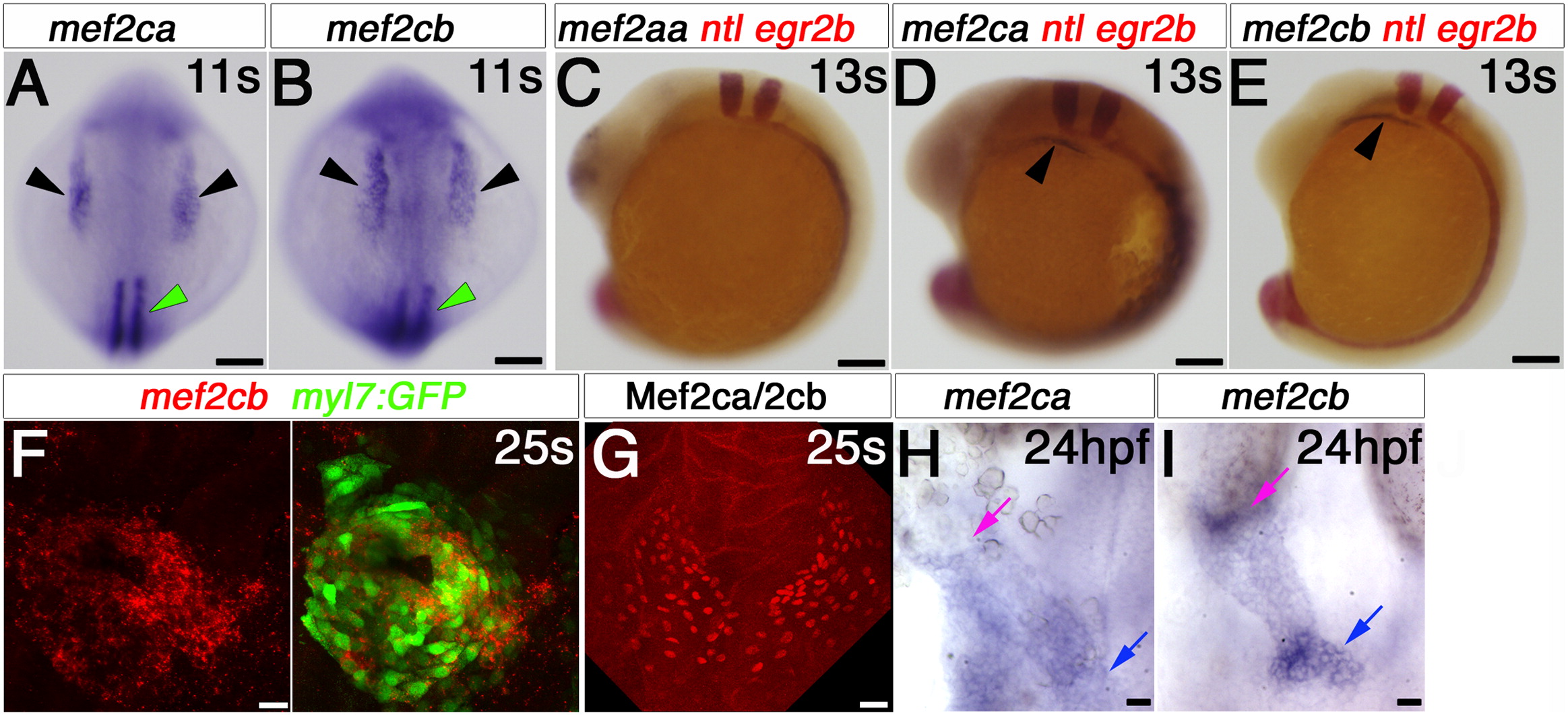
Fig. 1 Mef2ca and mef2cb expression during early cardiogenesis. In situ mRNA hybridisation of indicated genes, or immunodetection of Mef2ca/cb protein (G) for wild type (A–E, G–I) and Tg(myl7:EGFP) (F) in dorsal (A and B) or lateral (C–E) views of wholemount embryos or in flatmounts of dorsal views of the cardiac region, anterior to top (F–I). A and B. Mef2ca and mef2cb mRNAs accumulate in the bilateral heart fields in ALPM (black arrowheads) and adaxial cells (green arrowhead). C–E. Egr2b expression in rhombomeres 3 and 5, and ntl expression in the notochord positions the row of ventral cells in the ALPM (black arrowheads; D and E) that contain mef2ca and mef2cb, but not mef2aa mRNA (C). F. Confocal stack of Tg(myl7:EGFP) heart at 25 ss showing co-localisation of mef2cb mRNA (Fast Red) and EGFP. G. Mef2c protein in nuclei of a similar crescent of CMs spanning the midline. H and I. By 24 hpf, both mef2ca (H) and mef2cb (I) mRNAs are detected weakly in the heart tube, but mef2cb also accumulates strongly in the venous (blue arrow) and arterial (pink arrow) poles of the heart. Scale=100 μm (A–F), 20 μm (G–I). (For interpretation of the references to color in this figure legend, the reader is referred to the web version of this article.)
Reprinted from Developmental Biology, 369(2), Hinits, Y., Pan, L., Walker, C., Dowd, J., Moens, C.B., and Hughes, S.M., Zebrafish Mef2ca and Mef2cb are essential for both first and second heart field cardiomyocyte differentiation, 199-210, Copyright (2012) with permission from Elsevier. Full text @ Dev. Biol.