
|
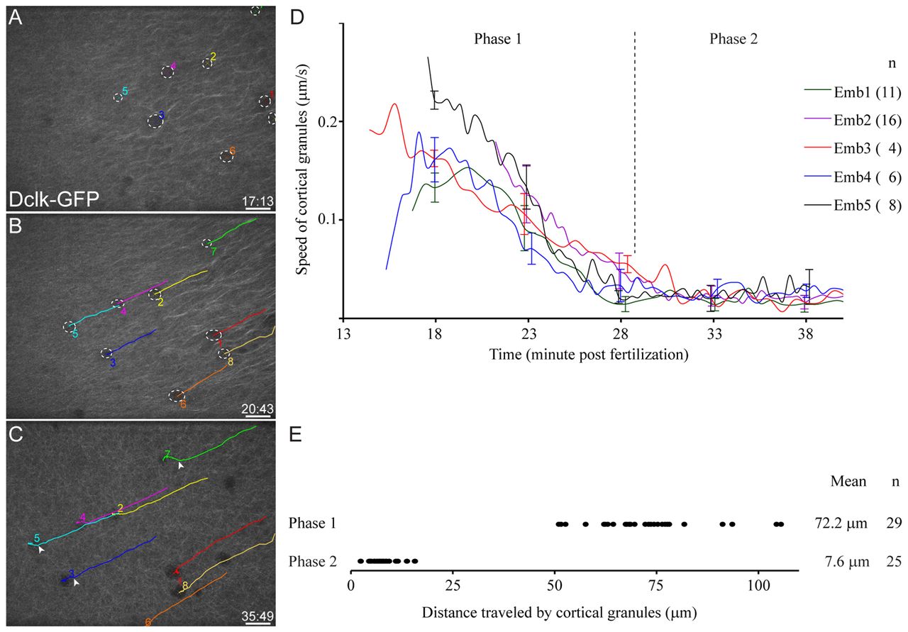
Fig. 3 Cortical granules at the vegetal pole are displaced by 20°. (A-D) Cortical granules (dotted white circles in A,B) at the vegetal pole move fast and in the same direction as parallel arrays before 30 mpf (Phase 1; B,D), and move slowly and randomly after parallel arrays disappear (Phase 2; C,D). Colored lines in B and C indicate individual cortical granules that were numbered and tracked. White arrowheads in C mark the position where cortical granules are no longer directional and begin moving randomly. (D) Each line shows the average speed of cortical granules in each embryo (number of granules tracked for each embryo shown in parentheses). Error bars are shown for selected time points and represent s.d. (E) Graph shows the distance traveled by individual cortical granules, and this measurement was used to calculate the angle of displacement. The cortical granules are displaced by ∼20°. Scale bars: 20 μm.