
|
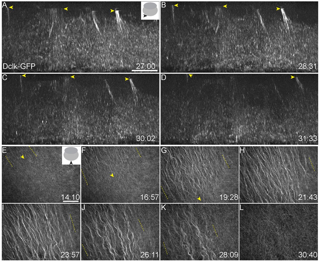
Fig. 1 Microtubule populations at the vegetal cortex of zebrafish embryos. (A-D) Tg(ef1α:dclk-GFP) expression shows perpendicular bundles at a depth of ∼15-30 μm inside the vegetal cortex, oriented along the animal-vegetal axis of the embryo. Yellow arrowheads indicate the position of perpendicular bundles moving through the yolk. (E-L) Parallel arrays form from ∼14 mpf, grow directionally and extend to cover a large area of the vegetal cortex before dissociating from 26 mpf. Parallel arrays are replaced by a non-directional meshwork (L) by ∼30 mpf. Yellow arrowheads (E-G) mark the front and yellow dashed lines (E-K) indicate the spread of parallel arrays. Insets in A and E show schematic of views, and viewing orientation is shown with solid black arrowheads. Numbers at bottom right of panels indicate mpf. Scale bars: in A, 10 μm; in E, 40 μm.