
|
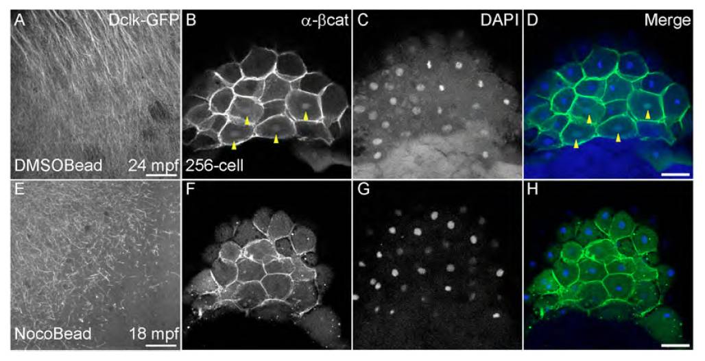
Fig. S3 Transient and local disruption of parallel microtubule arrays affects dorsal specification. (A,E) Dcklk-GFP labeled arrays shows local disruption in nocodazole bead-treated embryos (E) in comparison with control DMSO bead-treated embryos (A). (B,F) Nuclear accumulation of β-Catenin in DMSO bead-treated embryos (yellow arrowheads in B), in comparison with absence of β-Catenin in nocodazole bead-treated embryos (F). (C,D,G,H) DAPI staining (C,G) shows all nuclei in control and nocodazole-treated embryos, and D,H show merged images for β-Catenin and DAPI staining. Scale bars: in A,E, 30 μm; in D,H, 50 μm.