
Image
|
Figure Caption
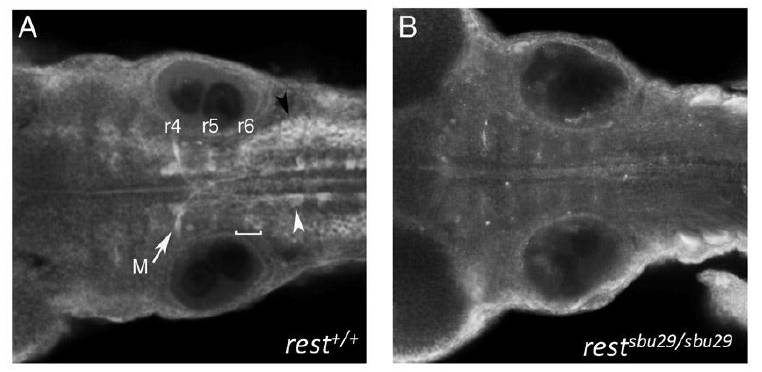
Fig. S4 Rest protein is expressed in FBMNs. Dorsal views of 48-hpf embryos, anterior to left. (A) Immunohistochemistry of wild-type embryos reveals Rest localization in reticulospinal neurons (white arrowhead), including the Mauthner (M) neuron (arrow), FBMNs (bracket), vagal motor neuron nuclei (black arrowhead), as well as in the surrounding neuroepithelium. (B) Staining of restsbu29/sbu29 homozygous mutant embryos does not detect Rest protein. Controls lacking the primary Rest antibody showed similar background fluorescence.
Acknowledgments
This image is the copyrighted work of the attributed author or publisher, and
ZFIN has permission only to display this image to its users.
Additional permissions should be obtained from the applicable author or publisher of the image.
Full text @ Development