
|
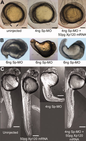
Fig. 5 p120 catenin δ1 depletion by δ1 splice-MO causes severe defects in somite formation in later stages of development including defects in yolk extension, tail extension, and eye formation. Embryos were imaged with Nomarski optics. A: Images of side views of live zebrafish embryos, anterior left, at the 14-somite stage (16 hpf). Embryos were uninjected or injected as indicated in the panels with δ1 splice-MO (Sp-MO) and/or Xenopus p120 catenin δ1 mRNA (Xp120 mRNA). Ventral arrows indicate the positions of the head and tailbud. Scale bars = 250 μm. B: Embryos were injected with 6 pg δ1 splice-MO (Sp-MO). C: Comparisons of side views of embryos that were uninjected or injected with δ1 splice-MO and/or Xp120 catenin δ1 mRNA at the 1-cell stage, and then imaged at the Prim 15 stage (30 hpf, anterior top). Eighty-six percent of the embryos injected with Sp-MO survived to Prim 15. Scale bars = 160 μm.