
|
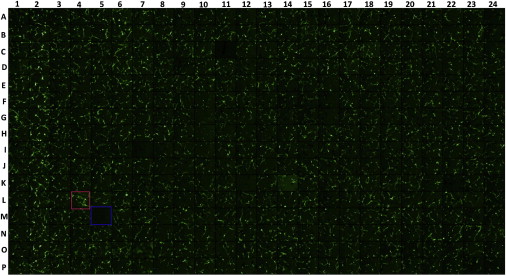
Fig. S4
Representative Image of flk1-GFP Endothelial Cells Growing in a 384-Well Plate at Day 5, Related to Figure 4 Column 2 is treated with 2 uM SB431542 as a positive control, and column 23 is treated with 1% DMSO as a negative control. Images of each well were recorded on a LHS-H100P-1 camera (Nikon, Japan) with the ImageXpressMICRO imaging system (Molecular Devices). Imaging DATA was processed with High Content Image Processing software and analyzed in the categories of total tube length, total tube area, and branch points with Angiogenesis Tube Formation (RD-1, MetaXpress, Molecular Devices). Red and blue squares show more and less tube formation, respectively.