
|
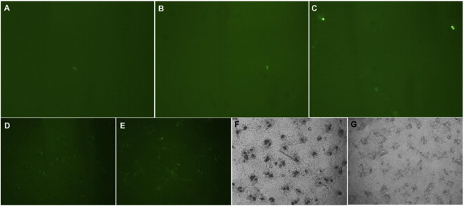
Fig. S2
Primary Cultures of Endoderm-Derived Insulin-GFP Pancreatic Beta Cells and Ectoderm-Derived vmat-2 GFP Neurons, Related to Figure 2 (A and B) Insulin-GFP cells are clearly detected at low frequency at day 1 (A) and day 2 (B) after culture. (C–F) Retinoic acid increases the number of insulin-GFP cells by <10-fold (C) compared with control (B). Vmat-2 GFP expression is observed at day 2 of culture (D). bFGF treatment at 100 ng/ml significantly increases GFP-positive cells and causes more neural network/branches (E) compared with control (D). Of interest, bFGF treatment (100 ng/ml) also remarkably inhibits the numbers of pigment cells (G) compared with control (F) at day 3.