
Image
|
Figure Caption
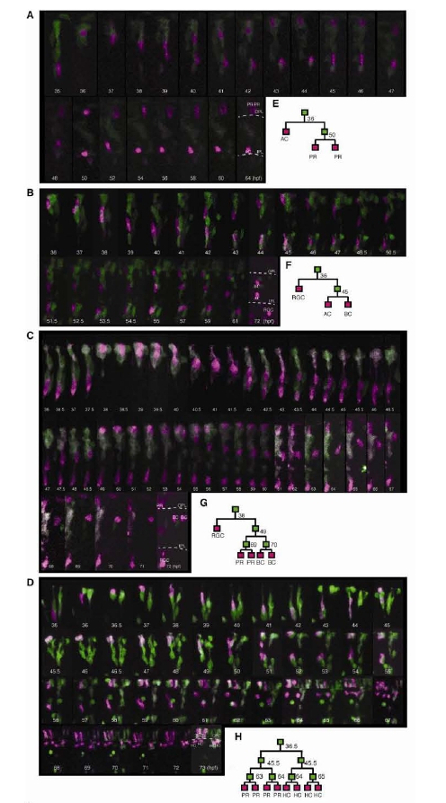
Fig. S4
Development of retinal clones derived from single RPCs photoconverted at 32 hpf. (A-D) Time series of two 3-cell, one 5-cell, one 8-cell clones generated from single RPCs (in Magenta), in which every division and cell fate are recorded (shown in the schematic reconstructed trees in (E-H). (I) Time lapse of retinal clones from 24 to 48 hpf. Shows reconstructed lineages from early (24-48 hpf) live imaging movies using MAZe fish. Dashed lines show Ihe clones which come from labelled RPCs that had already divided once by the time we began imaging.
Acknowledgments
This image is the copyrighted work of the attributed author or publisher, and
ZFIN has permission only to display this image to its users.
Additional permissions should be obtained from the applicable author or publisher of the image.
Full text @ Neuron