
|
Fig. S2
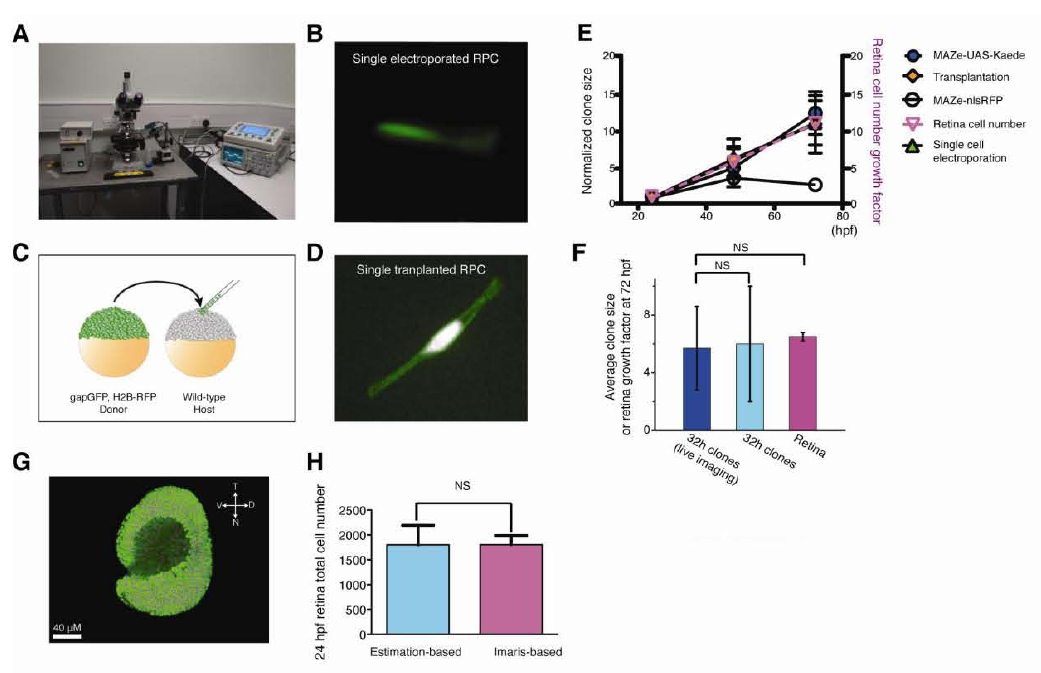
In vivo labeling of single retinal progenitor cells (RPCs). (A) A picture showing the setup for in vivo single-cell electroporation. (B) A single RPC electroporated with Dextran-Alexa 488 dye at 24 hpf. (C) A schematic diagram showing cell transplantation between a donor and host embryo. (D) Single RPC expressing gapGFP and H2B-RPF in a 24 hpf host retina following transplantation. (E) Plot showing that the clones generated by these different labeling methods grow in size in an indistinguished manner, which is further comparable to total cell number growth of retina tissue over development. However, significant cell loss was observed in MAZe-nlsRFP expressing clones, in which the average clone size decreases after 48hpf, finally dropping to 2.8 cells at 72 hpf(n=395), While, it is unclear why MAZe-nlsRFP labelled cells appear to die after 48hpf, this anomalous result emphasizes why it is critical to test the representativeness of clones induced by any labeling method if one is to conduct a rigorous clonal analysis of tissue development. (F) The size of 32 hpf-induced clones in live imaging is comparable to that of the 32h clones in the stain and fix experiment, and retina tissue growth. Values represent mean ± SD (n=71, 169, 4 for live imaging clones, 32h fixed clones and retina, NS, not significant). (G) Representative 3D reconstruction of a 24 hpf retina in which all the cell nucleus were recognized (Magenta color) by creating surface for every nucleus using Imaris. (H) Bar graph showing the total cell number of the 24 hpf retina measured by the cell number estimation protocol (left, Estimation-based) described in Method and the discontinuous surface-based approach (right, lmaris-based). Values represent mean ± SD (n=8, cell estimation-based protocol; n=4, Imaris-based method; NS, no significance; p > 0.05; Students′ t test). N: nasal; T: temporal; V: ventral; D: dorsal. All the cells express H2B-GFP.