
Image
|
Figure Caption
Fig. S1
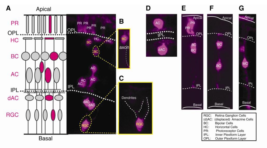
Retinal cell type identification. Distinct cell types are idcmified by the layer position and cell morphorlogy, which could be unambiguously examined in 3D reconstruction ofa clone, as shown in the Movie S I. (A) A schematic digram of the anatomical retinal structure (left) and a representative retinal clone expressing the photoconverted Kaede (right). (B,C) Zoomed Be and RGC in the clone (A, right). (D) An example oran AC and a displaced AC. (E) An example of two PRs and two BCs. (F) An example of two BCs. (G) An example of one Me.
Acknowledgments
This image is the copyrighted work of the attributed author or publisher, and
ZFIN has permission only to display this image to its users.
Additional permissions should be obtained from the applicable author or publisher of the image.
Full text @ Neuron