
|
Fig. S4
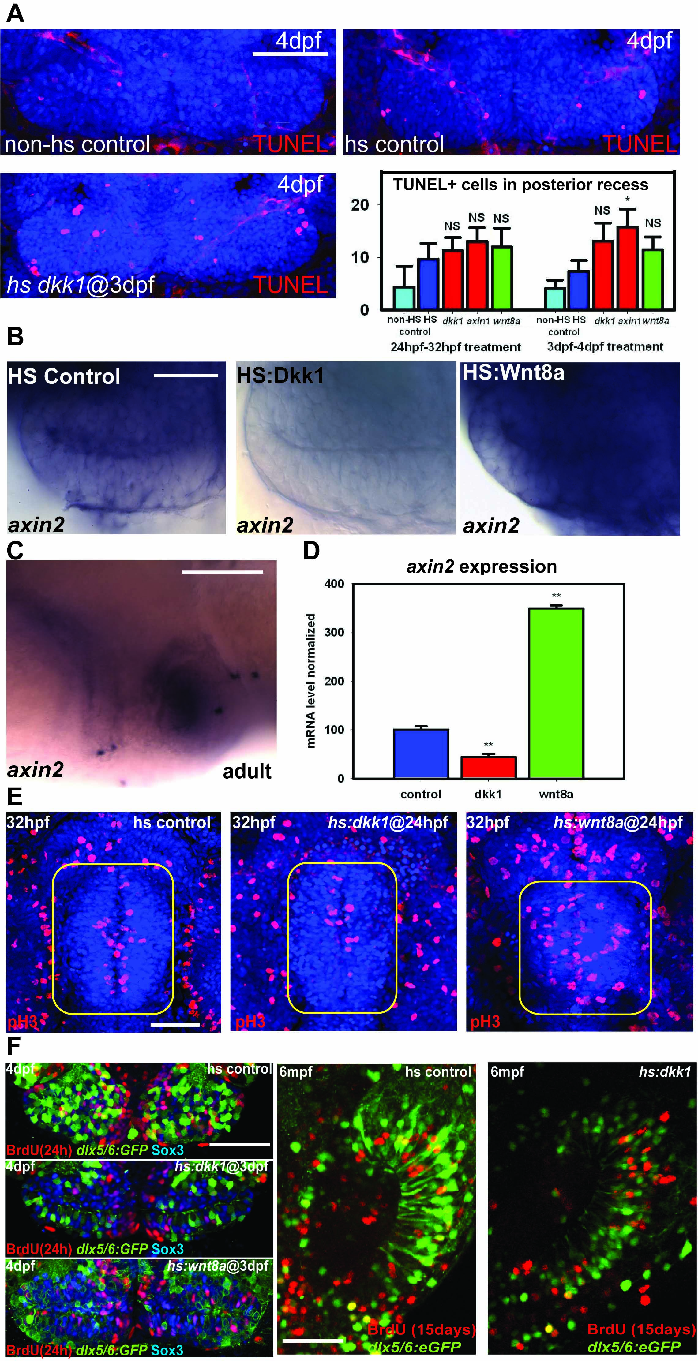
Effects of Wnt signaling on hypothalamic progenitors, related to Figure 3. (A) Ventral confocal optical sections of TUNEL staining at 4dpf, and counts of TUNEL+ cells at 32hpf and 4dpf. Only hs:axin1 expression from 3-4dpf significantly increases cell death compared to heat shock alone. (B) axin2 in situ hybridization at 4dpf following heat shock at 3dpf. (C) axin2 in situ hybridization in a mid-sagittal section of the adult hypothalamus. Red oval marks posterior recess. (D) axin2 mRNA expression levels in the dissected adult hypothalamus following Wnt pathway inhibition or activation for 15 days. (E) Phospho-Histone H3 staining in the 32hpf hypothalamus (yellow squares). (F) Co-localization of BrdU labeling with Sox3 and dlx5/6:GFP labeling in the 4dpf and adult posterior recess. Single confocal optical sections are shown in all panels. Scale bars: (A,C,F) 80μm, (B) 25μm, (E) 50μm. Cell counts were collected from ventral maximum intensity confocal Z-projections through 3 individual brains. The entire hypothalamus was counted at 32hpf, and the entire posterior recess was counted at 4dpf. **: p<0.05 compared to non-HS control. Error=±SD.
Reprinted from Developmental Cell, 23(3), Wang, X., Kopinke, D., Lin, J., McPherson, A.D., Duncan, R.N., Otsuna, H., Moro, E., Hoshijima, K., Grunwald, D.J., Argenton, F., Chien, C.B., Murtaugh, L.C., and Dorsky, R.I., Wnt signaling regulates postembryonic hypothalamic progenitor differentiation, 624-636, Copyright (2012) with permission from Elsevier. Full text @ Dev. Cell