
|
Fig. S3
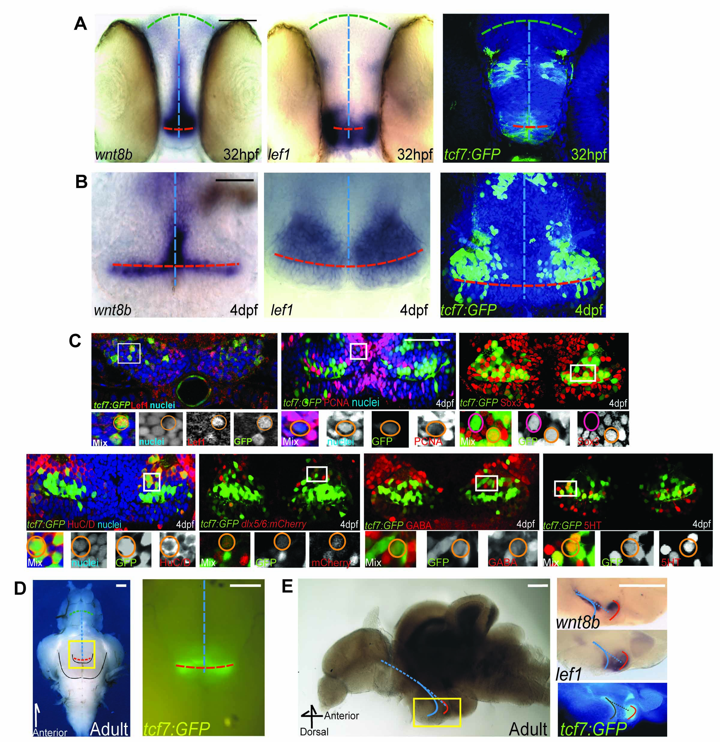
Expression of Wnt pathway components in zebrafish hypothalamic neural progenitors, related to Figure 2. (A,B) wnt8b mRNA, lef1 mRNA, and tcf7:GFP expression at 32hpf (A), and 4dpf (B) in ventral whole-mount views. (C) Co-expression of tcf7:GFP in the 4dpf hypothalamus with cell-type specific markers. (D) Bright-field and fluorescent dissecting microscope ventral images of the adult hypothalamus expressing tcf7:GFP. Yellow box in the bright field image indicates the region magnified in the fluorescent image. (E) Mid-sagittal view of wnt8b mRNA, lef1 mRNA, and tcf7:GFP expression in the adult hypothalamus. Yellow box in the left image indicates the region magnified in the right images. Blue line: 3rd ventricle; Red line: Posterior recess. A 12μm transverse cryosection is shown for Lef1 staining in (C), all other panels show single confocal optical sections from ventral views unless otherwise indicated. Scale bars: (A,B) 50μm, (C) 80μm, (D,E) 250μm.
Reprinted from Developmental Cell, 23(3), Wang, X., Kopinke, D., Lin, J., McPherson, A.D., Duncan, R.N., Otsuna, H., Moro, E., Hoshijima, K., Grunwald, D.J., Argenton, F., Chien, C.B., Murtaugh, L.C., and Dorsky, R.I., Wnt signaling regulates postembryonic hypothalamic progenitor differentiation, 624-636, Copyright (2012) with permission from Elsevier. Full text @ Dev. Cell