
|
Fig. S2
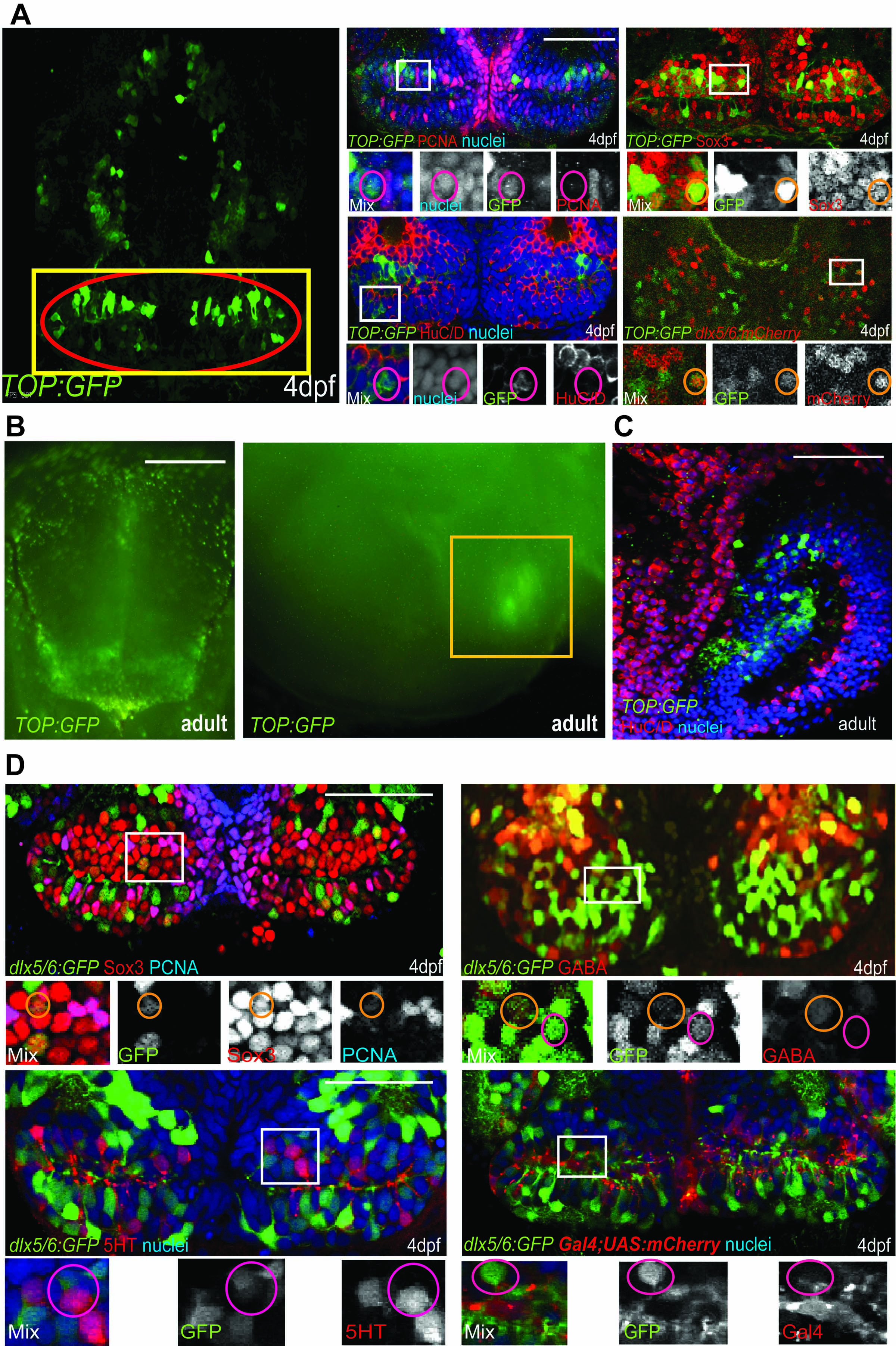
Identity of reporter-labeled cells, related to Table 1. (A) Maximum-intensity confocal ventral projection of 4dpf TOP:GFP hypothalamus on left, yellow box marks the area shown on right and red oval marks the posterior recess. Co-staining of GFP with cell-type specific markers is shown on right. (B) Dissecting microscope images of TOP:GFP expression in the adult hypothalamus, from ventral and mid-sagittal views. Yellow box marks the area shown in (C). (C) Co-staining of GFP and HuC/D in the adult hypothalamus. (D) dlx5/6:GFP expression overlaps with Sox3, PCNA, and GABA, but not with 5HT or a Gal4 insertion expressed in radial glia (C). White boxes indicate enlarged regions. Small orange circles label cells with colocalization and small magenta circles label cells without colocalization. Single confocal optical sections are shown unless otherwise indicated. Scale bars: Scale bars: (A,C,D) 80μm, (B) 250μm.
Reprinted from Developmental Cell, 23(3), Wang, X., Kopinke, D., Lin, J., McPherson, A.D., Duncan, R.N., Otsuna, H., Moro, E., Hoshijima, K., Grunwald, D.J., Argenton, F., Chien, C.B., Murtaugh, L.C., and Dorsky, R.I., Wnt signaling regulates postembryonic hypothalamic progenitor differentiation, 624-636, Copyright (2012) with permission from Elsevier. Full text @ Dev. Cell