
|
Fig. S1
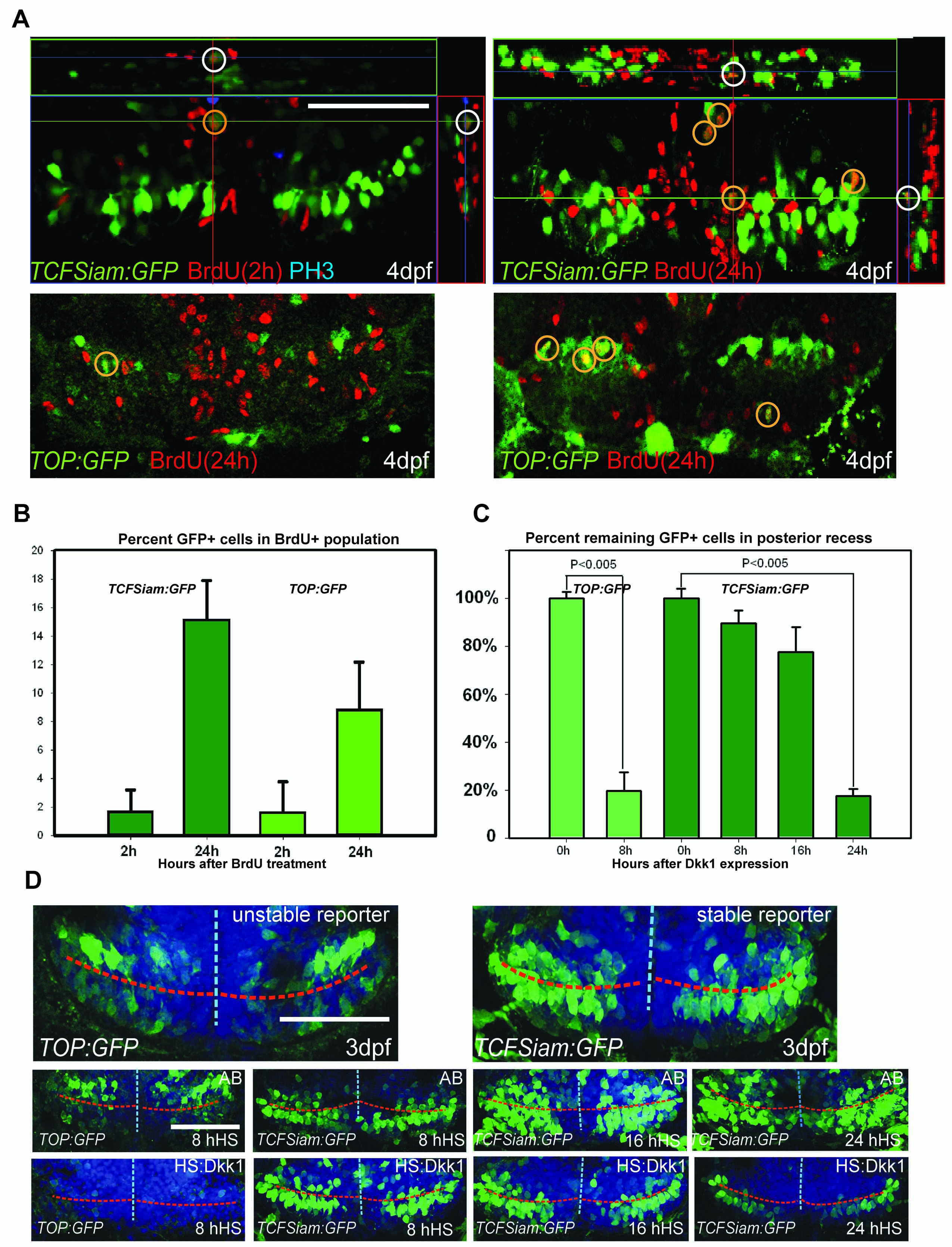
Analysis of Wnt reporter at 4dpf, related to Figure 1. (A) Ventral view of the posterior recess region of TOP:GFP and TCFSiam:GFP hypothalamus after short (2h) and long term (24h) BrdU labeling; observed with 50μm maximum intensity confocal Z-projections. (B) Quantification of BrdU+/GFP+ cells 2 hours or 24 hours after labeling. (C) Relative percentages of GFP+ cells following hs:dkk1 expression at 3dpf, compared to wild-type controls. (D) Ventral view of the posterior recess region of TOP:GFP and TCFSiam:GFP embryos, 8h, 16h, and 24h after hs:dkk1 activation at 3dpf. Images are 50μm ventral maximum intensity confocal Z-projections. Scale bars: 80μm. Cell counts were collected from ventral maximum intensity confocal Z-projections through the posterior recess of 3 individual samples for each condition. Error=±SD.
Reprinted from Developmental Cell, 23(3), Wang, X., Kopinke, D., Lin, J., McPherson, A.D., Duncan, R.N., Otsuna, H., Moro, E., Hoshijima, K., Grunwald, D.J., Argenton, F., Chien, C.B., Murtaugh, L.C., and Dorsky, R.I., Wnt signaling regulates postembryonic hypothalamic progenitor differentiation, 624-636, Copyright (2012) with permission from Elsevier. Full text @ Dev. Cell