
|
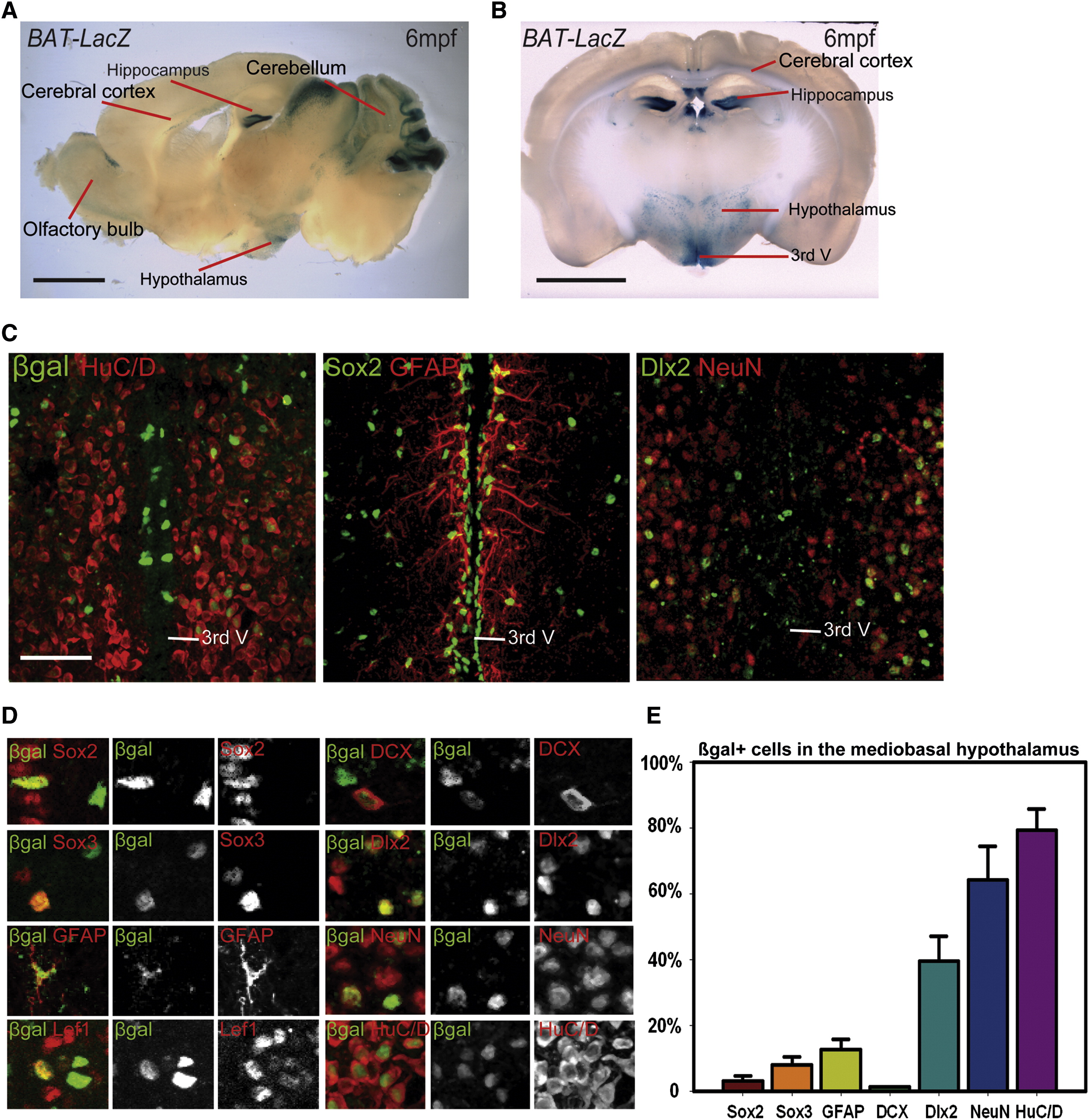
Fig. 5
The Adult Mouse Hypothalamus Has a Wnt-Responsive Cell Population (A and B) Sagittal (A) and coronal (B) sections of adult Bat-LacZ mouse brains. (C) β-gal+ cells are distributed in both the ventricular zone (where Sox2+ and GFAP+ cells reside) and the parenchymal zone (where Sox2, HuC/D+, NeuN+, and Dlx2+ cells reside) Coronal 40μm cryosections are shown. (D) Colocalization of β-gal with specific markers in the hypothalamus. (E) Percentage of marker coexpression within the β-gal+ population. Single confocal optical sections are shown in (C) and (D). Scale bars represent 2 mm (A and B), and 80 μm (C). Cell counts were collected from the mediobasal hypothalamus, using six 40μm cryosections each from three mice. Error = ±SD.
Reprinted from Developmental Cell, 23(3), Wang, X., Kopinke, D., Lin, J., McPherson, A.D., Duncan, R.N., Otsuna, H., Moro, E., Hoshijima, K., Grunwald, D.J., Argenton, F., Chien, C.B., Murtaugh, L.C., and Dorsky, R.I., Wnt signaling regulates postembryonic hypothalamic progenitor differentiation, 624-636, Copyright (2012) with permission from Elsevier. Full text @ Dev. Cell