
|
Fig. 5
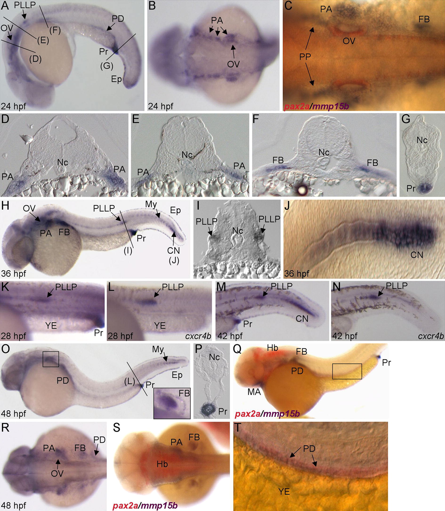
Expression pattern of mmp15b during pharyngula and hatching periods. (A) Lateral view of 24 hpf embryo with anterior to the left. Black lines denote cross-section slices shown in panels (D–G). (B and C) Dorsal views of 24 hpf embryos with anterior to the left. Embryo in panel (C) is double-labeled with mmp15b and pax2a (red). (D–G) Cross-sections of 24 hpf embryos at positions marked in panel (A). (H and J) Lateral views of 36 hpf embryos with anterior to the left. (I) Cross-section of 36 hpf embryo trunk (position shown in panel H) showing expression in posterior lateral line primordia, PLLP. Mmp15b and cxcr4b expression at 28 hpf (K and L) and 42 hpf (M and N), lateral views with anterior to the left and dorsal to the top. YE, yolk extension. (O, Q, and T) Lateral views of 48 hpf embryos with anterior to the left. Inset in panel (O) shows close-up of mmp15b expression in the fin bud, FB. (P) Cross-section through the proctodeum (Pr) of a 48 hpf embryo (position shown in panel O). (R and S) Dorsal views of 48 hpf embryos with anterior to the left. (Q, S, and T) Double labeling with pax2a (red) highlights mmp15b expression in pharyngeal arches (PA) and pronephric duct, PD. CN, caudal notochord; Ep, epidermis; Hb, hindbrain; MA, mandibular arch; My, myoseptum; Nc, notochord; OV, otic vesicle; PP, pharyngeal pouch.
Reprinted from Gene expression patterns : GEP, 12(7-8), Quick, R.E., Dunlap, J.A., and Jessen, J.R., Expression analysis of zebrafish membrane type-2 matrix metalloproteinases during embryonic development, 254-260, Copyright (2012) with permission from Elsevier. Full text @ Gene Expr. Patterns