
|
Fig. S1
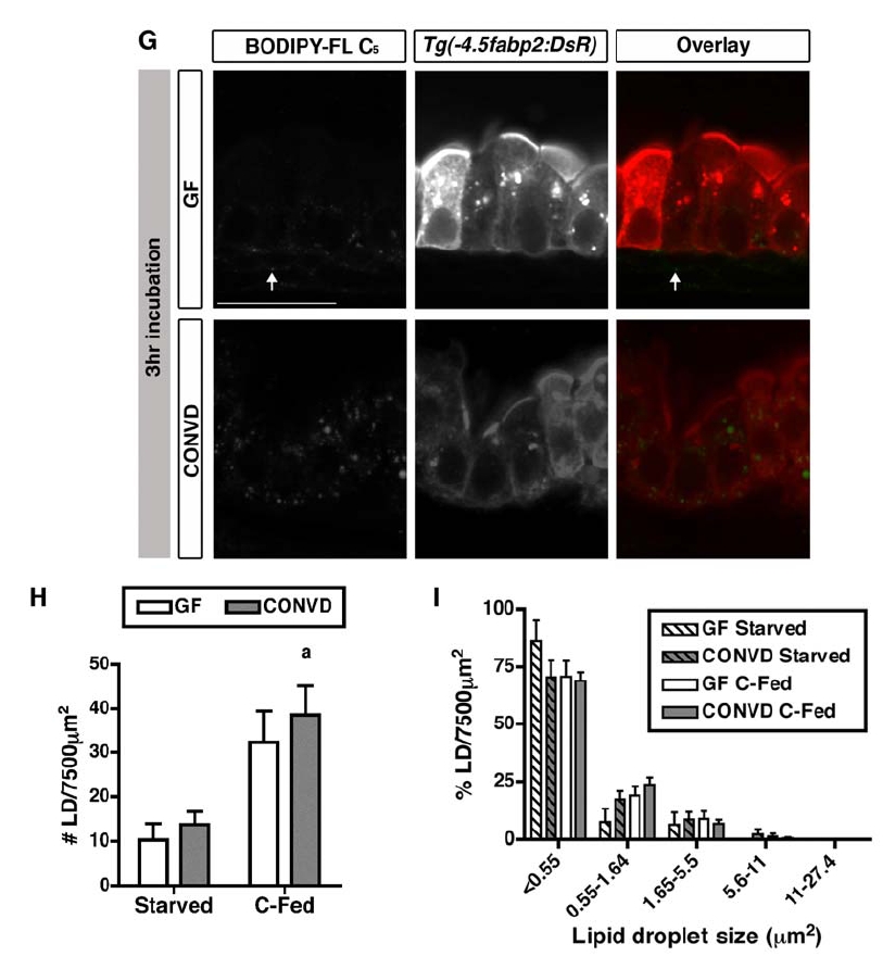
related to Figure 2. Lipid Droplet Quantification Assay Reveals Similar Lipid Droplet Formation in 6 dpf GF and CONVD Zebrafish Incubated with BODIPY-FL C5 Liposomes for 3 hr
(A) Z-stack movies collected on the Olympus FV1000 laser scanning microscope were scanned using the FV1000 FLUOVIEW software. To determine single epithelial layers, we overlayed the GFP signal from the BODIPY-C5 fatty acid and the DsRed signal from the Tg(-4.5fabp2:DsRed) line, which expresses DsRed in intestinal epithelial cells. We selected three slices with independent regions of interest per fish and extracted the GFP signal only in Volocity 5.5.1. (BF) LD number and size quantification in Volocity. We imported the Z-stacks from individual fish in Volocity and opened the selected slices (B) to quantify the fluorescent signal. (C) Scale arrow, 20 μm. The red box shows the magnified region in D and E. After the region of interest was selected (purple box in D), we applied our quantification protocol (F) to identify individual LDs as objects (E,F) with displayed size (Area, μm2). (F) Scale arrow, 45 μm.The selected slice shows a medium (green arrow) and a large LD (black arrowhead, E,F). (G) Representative confocal images of fixed 6 dpf Tg(-4.5fabp2:DsRed) GF and CONVD zebrafish fed a control diet and incubated with BODIPY-FL C5 for 3 hr. The lumen is at the top and epithelium at the bottom of all images. Scale bar, 20 μm. Intestinal epithelial cells are identified by DsRed expression, and BODIPY-C5 is detected in the epithelium as small LDs and in the lamina propria (arrow; A). (H and I) The LD quantification assay described above was used to determine LD number (H) and relative size frequency (I) in an epithelial region of interest (7500 μm2). The graphs represent the mean ± SEM in starved and control-fed (C-Fed) zebrafish (at least two independent experiments; 4-10 fish/condition/experiment). Results of Student’s t test corrected.