
|
Fig. 2
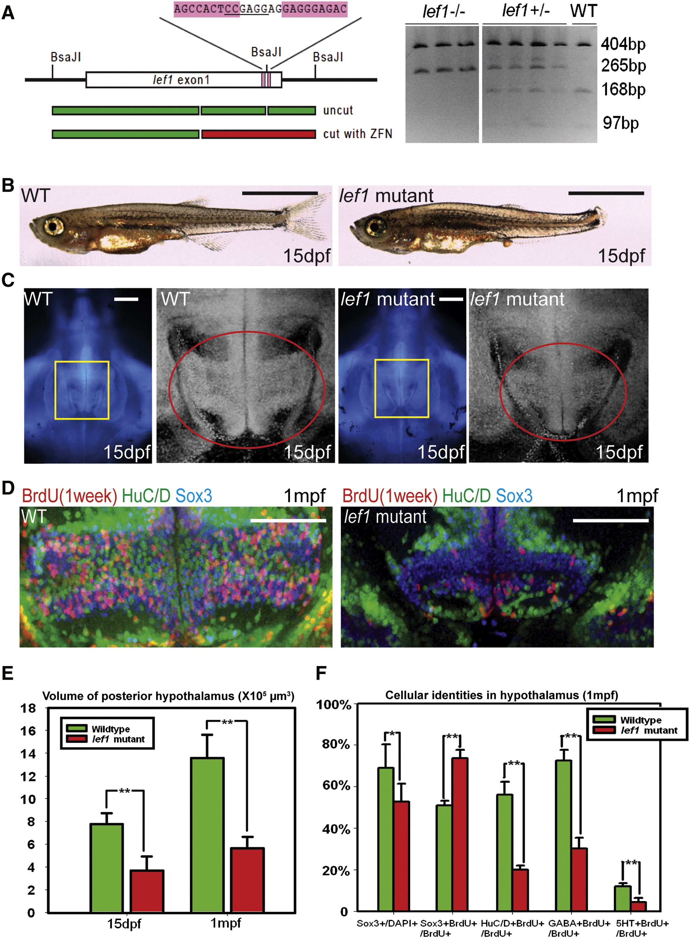
lef1 Is Required for Proliferation and Neurogenesis in the Postembryonic Hypothalamus (A) lef1 ZFN target region and genotyping. (B and C) Whole fish (B) and brain size (C) comparisons of lef1 mutant to wild-type at 15 dpf. Boxed region in left panel is enlarged on right, posterior recess is circled. (D) Expression of HuC/D, Sox3, and 7-day BrdU labeling in posterior recess of wild-type and lef1 mutant hypothalamus. (E) Quantification of posterior hypothalamus size. (F) Tracing of proliferating cells. lef1 mutants have a smaller Sox3+ progenitor pool, but a higher percentage of BrdU+ cells express Sox3 and fewer produce HuC/D+, GABA+, or 5HT+ neurons. Single optical sections from ventral views are shown in (C) and (D). Scale bars represent 2 mm (B), 200 μm (C), and 100 μm (D). Brain volumes were calculated using Amira software. Cell counts were collected from maximum intensity Z projections through the posterior recess of three individual samples for each genotype and calculated using Volocity software. *p < 0.05, **p < 0.005. Error = ±SD. See also Figure S3.
Reprinted from Developmental Cell, 23(3), Wang, X., Kopinke, D., Lin, J., McPherson, A.D., Duncan, R.N., Otsuna, H., Moro, E., Hoshijima, K., Grunwald, D.J., Argenton, F., Chien, C.B., Murtaugh, L.C., and Dorsky, R.I., Wnt signaling regulates postembryonic hypothalamic progenitor differentiation, 624-636, Copyright (2012) with permission from Elsevier. Full text @ Dev. Cell