
|
Fig. 1
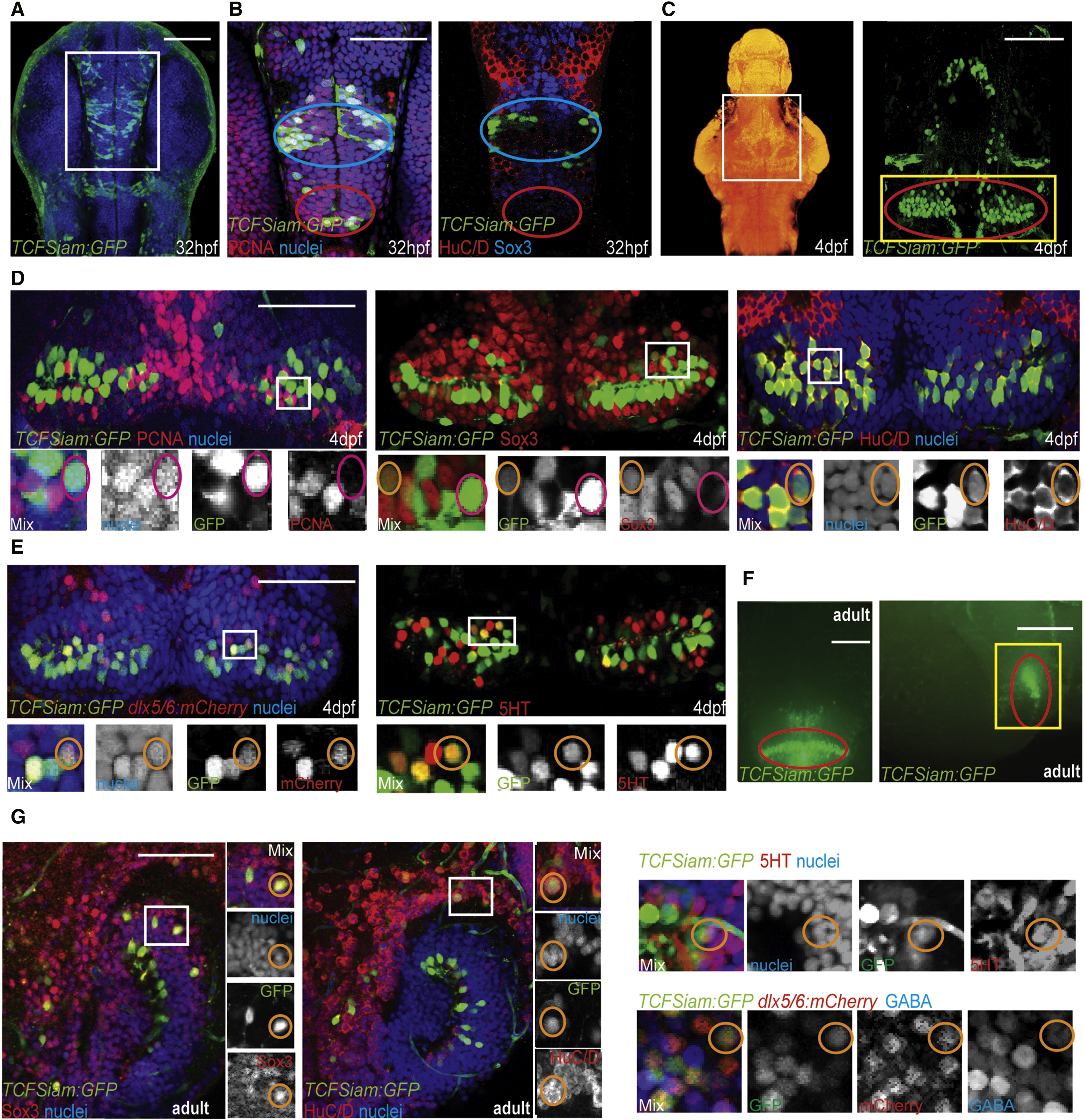
Identification of Wnt-Responsive Cells in the Zebrafish Hypothalamus (A) Ventral view of TCFSiam:GFP. (B) Costaining with PCNA, Sox3, and Hu. Blue ovals label the presumptive lateral recess, and red ovals label the presumptive posterior recess. White box in (A) marks the area shown. (C) Maximum intensity confocal Z-projection of TCFSiam:GFP at 4 dpf. In nuclear stained ventral view of brain on left, white box marks hypothalamic area shown on right. Red oval marks the posterior recess. (D) Costaining of TCFSiam:GFP with PCNA, Sox3, and HuC/D. Yellow box in (C) marks the area shown. (E) Costaining of TCFSiam:GFP with dlx5/6:gfp and serotonin. Yellow box in (C) marks the area shown. (F) Dissecting microscope ventral and sagittal views of TCFSiam:GFP in the adult hypothalamus. Red oval labels the presumptive posterior recess. (G) Costaining of TCFSiam:GFP with Sox3, Hu, 5HT, dlx5/6:mCherry, and GABA in the adult posterior hypothalamus. Yellow box in (F) marks the area shown. Single optical sections from ventral views are shown in all panels, unless otherwise indicated. Small orange circles label cells with colocalization, and small magenta circles label cells without colocalization. Scale bars represent 80 μm (A–E,G), and 250 μm (F). See also Figure S1.
Reprinted from Developmental Cell, 23(3), Wang, X., Kopinke, D., Lin, J., McPherson, A.D., Duncan, R.N., Otsuna, H., Moro, E., Hoshijima, K., Grunwald, D.J., Argenton, F., Chien, C.B., Murtaugh, L.C., and Dorsky, R.I., Wnt signaling regulates postembryonic hypothalamic progenitor differentiation, 624-636, Copyright (2012) with permission from Elsevier. Full text @ Dev. Cell