
|
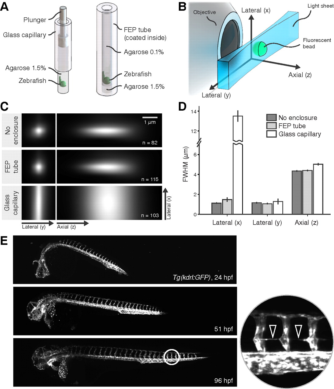
Fig. 3 Optical quality of FEP mounting. (A) Schematic of mounting in 1.5% agarose (left) and multilayer mounting in 0.1% agarose in coated FEP tubes (right). (B) Principle of SPIM and direction of axes for the analysis of optical quality. Not to scale. (C) Maximum intensity projections of averaged images of beads (n, number of beads) in 1.5% agarose without enclosure, in an FEP tube and in a glass capillary. (D) Full width at half maximum (FWHM). Mean ± s.d., n=6. (E) Developing zebrafish embryo [Tg(kdrl:GFP)] embedded in 0.1% agarose in a coated FEP tube during SPIM time-lapse (supplementary material Movie 1). Images resolve even the finest structures, e.g. the cell extensions forming the parachordal vessels (arrowheads).