
Image
|
Figure Caption
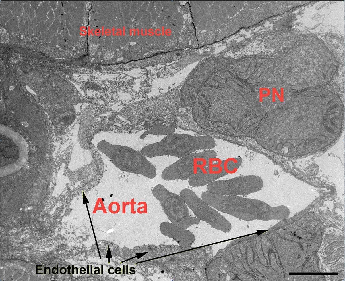
Fig. S2 Transmission electron microscopy shows that the trunk region of the dorsal aorta of the zebrafish embryo was devoid of smooth muscle cells. This image, taken from the midpoint of the region of the aorta used for time based imaging, demonstrates that no smooth muscle cells are present around the vessel at 5 dpf. Skeletal muscle cells are clearly visible, as is the pronephros (PN). Erythrocytes (RBC) are abundant within the vessel, and endothelial cells are clearly evident. No smooth muscle cells can be detected. (Scale Bar = 20 microns).
Acknowledgments
This image is the copyrighted work of the attributed author or publisher, and
ZFIN has permission only to display this image to its users.
Additional permissions should be obtained from the applicable author or publisher of the image.
Full text @ PLoS One