
|
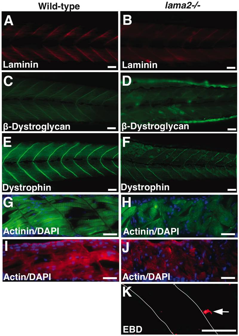
Fig. 3 Laminin-α2 deficiency results in severe muscle degeneration.
(A–B) Wholemount immunofluorescence analysis showed reduced levels of laminin complex in myotendinous junctions in the mutant muscles. (C–F) The expression of β-dystroglycan as well as dystrophin was also reduced in the mutant muscles. (G–J) Myofibers in wild-type muscles are attached to either side of the myotendinous junctions and displayed well-organized muscles. Mutant fish, however, displayed highly disorganized muscles. Several detached myofibers lacking the contractile proteins are seen in the mutant muscles by α-actinin and sarcomeric actin antibody staining (arrows). (K) Evans blue dye (EBD) injections at 3 dpf detected occasional staining in necrotic fibers (arrow). Bars = 10 μm.