
|
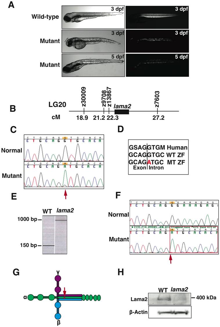
Fig. 1 Morphological, genetic and molecular characterization of lama2cl501/cl501 zebrafish.
(A) In comparison to the wild-type fish, lama2cl501/cl501 fish exhibit smaller bodies (left panel) and displayed muscle disorganization early in development at 3 dpf (right panel). The muscle degeneration progressed to most of the somites by 5 dpf. (B) Genetic mapping in mutant fish associated the linkage of mutant locus in 4.9 cM region on chromosome 20 in zebrafish identifying lama2 as the candidate gene. (C) Sequencing of lama2 gene in zebrafish identified a mutation at c.5376+1G>A in the mutant fish (arrow). (D) The consensus splice site containing the mutation is highly conserved in human LAMA2 gene. (E) RT-PCR using primers specific for the exons 33 and 34 on either side of the mutant intronic sequence showed a larger PCR product (~900 base pairs) in the mutant fish in comparison to the wild-type fish (152 base pairs) suggesting aberrant splicing. (F) Sequencing of the RT-PCR products revealed the retention of 764 base pairs of intron 33 in the mutation fish (arrow). (G) This mutation is localized at the long arm coiled-coil of lama2 protein (arrow) that interacts with β- and γ- laminins to form functional laminin complex (H) Western blotting analysis showed a reduction of laminin-α2 protein in the mutant fish.