
|
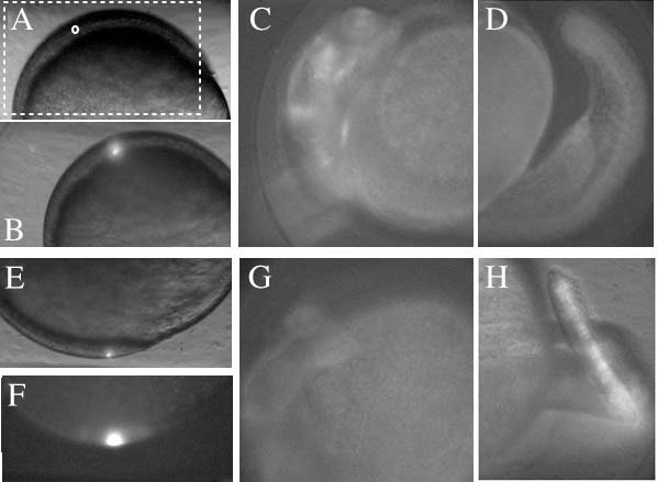
Fig. S5
Local labeling with photo-activation of Kaede. Local illumination with a UV laser at ~80% epiboly in the precursor of the head region. To confirm the localization of the UV laser illumination we used the photoconversion of Kaede (from green to red fluorescence) to track the fate of the illuminated region. The mRNA encoding Kaede (Kohei et al., 2006) was injected at the one-cell stage. (A) Embryo overview. The boxed region indicating the anterior part of the embryo corresponds to the area shown in B,C. The region indicated by the small circle was selected for UV irradiation. (B) Twenty seconds of UV irradiation (bright spot) in the circled region in A. (C,D) At 24 hpf, red fluorescence from diffused Kaede was observed in the head (C), but not in the tail (D). (E) UV irradiation for 20 seconds in a region (bright spot) that develops into the tail. (F) Corresponding red fluorescent image immediately after photoconversion. (G,H) At 24 hpf, red fluorescence was observed in the tail (H) but not in the head (G).