
|
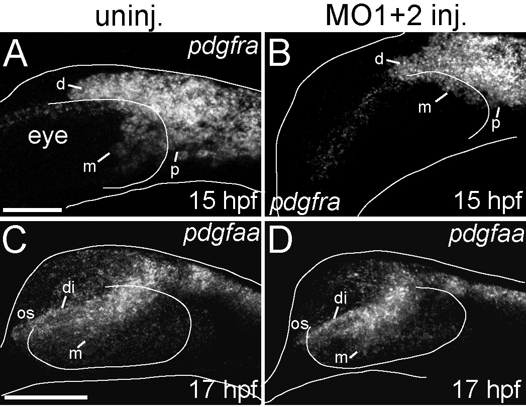
Fig. 4
pdgframRNA expression is down-regulated inhdac4MO-injected embryos, although the ligandpdgfaais unaffected. (A-D) mRNA in situ hybridizations, lateral views where anterior is towards the left, dorsal is upwards, Images are projections from confocal stacks. Dorsal and ventral margins and the eye margin of the embryo were outlined from brightfield images. (A) Uninjected embryos showed expression of pdgfra dorsal to the eye (d), posterior to the eye (p), and medial to the eye (m) at 15 hpf. (B) MO-injected embryos showed more limited expression of pdgfra, in particular loss of pdgfra expression medial to the eye (m) at 15 hpf (compare m in A with B). (C) At 17 hpf, uninjected embryos showed pdgfaa mRNA expression in the diencephalon (di), medial to the eye (m), and in the optic stalk anterior to the eye (os). (D) At 17 hpf, hdac4 MO-injected embryos had similar patterns of pdgfaa mRNA expression in the diencephalon (di), medial to the eye (m), and in the optic stalk (os). A and B, scale bar = 50 μm, C-D scale bar = 100 μm.