
|
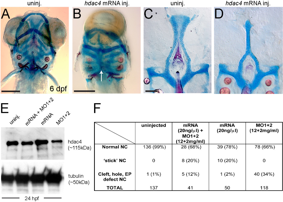
Fig. 3
Over-expression ofhdac4mRNA rescues palatal defects associated with MO-injection and causes midline craniofacial defects. (A-D) Alcian Blue (cartilage) and Alizarin Red (bone) stained embryos and dissected palatal skeletons. (A and B) Ventral view of whole-mount skeletal preparations of 6 dpf larvae, anterior is upwards, ventral is facing. (C and D) Flat-mounts of palatal skeletons at 6 dpf. Anterior is upwards. (A) Uninjected fish have normal development of craniofacial cartilage and bone. (B) hdac4 mRNA injection resulted in cyclopia and a loss of normal midline patterning (indicated by white arrow). (C) Uninjected fish have normal palatal skeletons. (D) hdac4 mRNA injection results in ‘stick’-like palatal skeletons. (E) Co-injection of hdac4 mRNA (20 ng/µl) and MO1 + 2 (12 + 2 mg/ml) resulted in protein levels similar to levels in uninjected embryos 24 hours post-injection, compared with injection of mRNA or MOs alone. (F) Embryos used for the protein assay were also raised to 6 dpf and scored for palatal phenotypes. Co-injection of hdac4 mRNA with MO1 + 2 resulted in a decrease of MO-like defects (i.e. cleft, hole, ethmoid plate (EP) defects) compared with MO-injection alone. A and B, scale bar = 200 μm; C and D, scale bar = 100 μm.