
Image
|
Figure Caption
Fig. S5
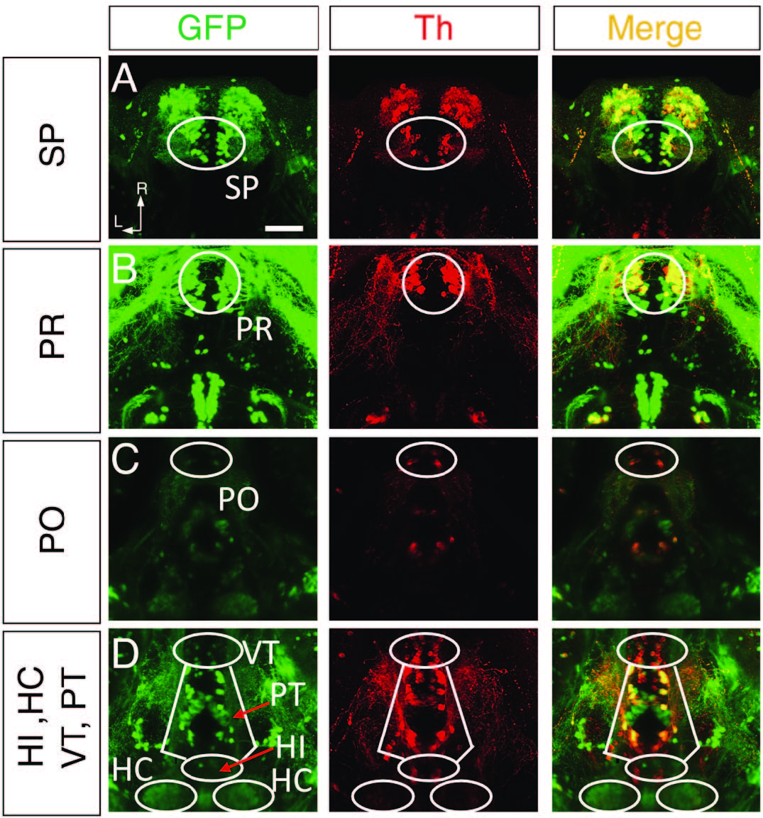
related to Figure 7. Tyrosine Hydroxylase (Th)-Immunostaining of ETvmat2:GFP Larvae
(A-D) Projected confocal images showing GFP-immunoreactivity (GFP-ir,left), Th-immunoreactivity (Th-ir, middle) and merged (right) signals in the subpallium (SP), pretectum (PR), preoptic area (PO), ventral thalamus (VT), posterior tubercular (PT),intermediate hypothalamus (HI) and caudal hypothalamus (HC). Each cluster is delineated by white lines. The images were obtained from the same larva aged at 4 dpf. The z-stack movies are shown in Movies S4-S5. Dorsal view, rostral is up. L, lateral; R, rostral. Scale: 50 μm.
Figure Data
Acknowledgments
This image is the copyrighted work of the attributed author or publisher, and
ZFIN has permission only to display this image to its users.
Additional permissions should be obtained from the applicable author or publisher of the image.
Full text @ Neuron