
|
Fig. S2
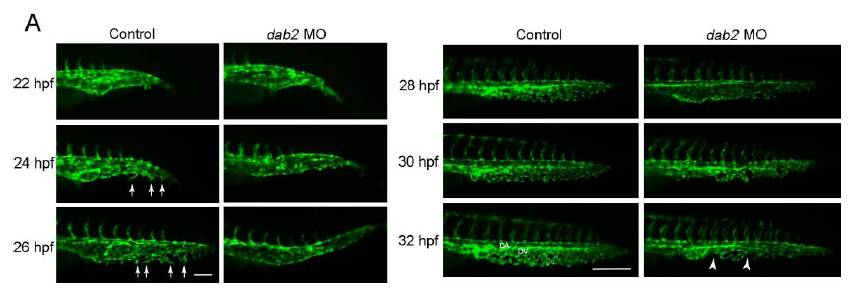
CVP formation in wild-type and dab2 deficient embryos.
(A) CVP development in dab2 MO injected embryos. In dab2 MO injected embryos, ventral sprouts from the caudal vein failed to emerge at 22hpf. Until 28hpf, the developing CVP in dab2 MO injected embryos are largely devoid of ventral sprouts. This initial defect in the formation of ventral sprouts caused dysmorphic CVP at 32hpf in dab2 MO injected embryos. Arrows present ventral sprouting of endothelial cells and arrowheads point to gaps within the CVP. Abbreviations: DA: Dorsal Aorta, DV: Dorsal Vein, VV: Ventral Vein. Scale bars are 100μm for 26hpf and 200μm for 32hpf.
Reprinted from Developmental Cell, 23(2), Kim, J.D., Kang, H., Larrivée, B., Lee, M.Y., Mettlen, M., Schmid, S.L., Roman, B.L., Qyang, Y., Eichmann, A., and Jin, S.W., Context-dependent proangiogenic function of bone morphogenetic protein signaling is mediated by disabled homolog 2, 441-448, Copyright (2012) with permission from Elsevier. Full text @ Dev. Cell