
|
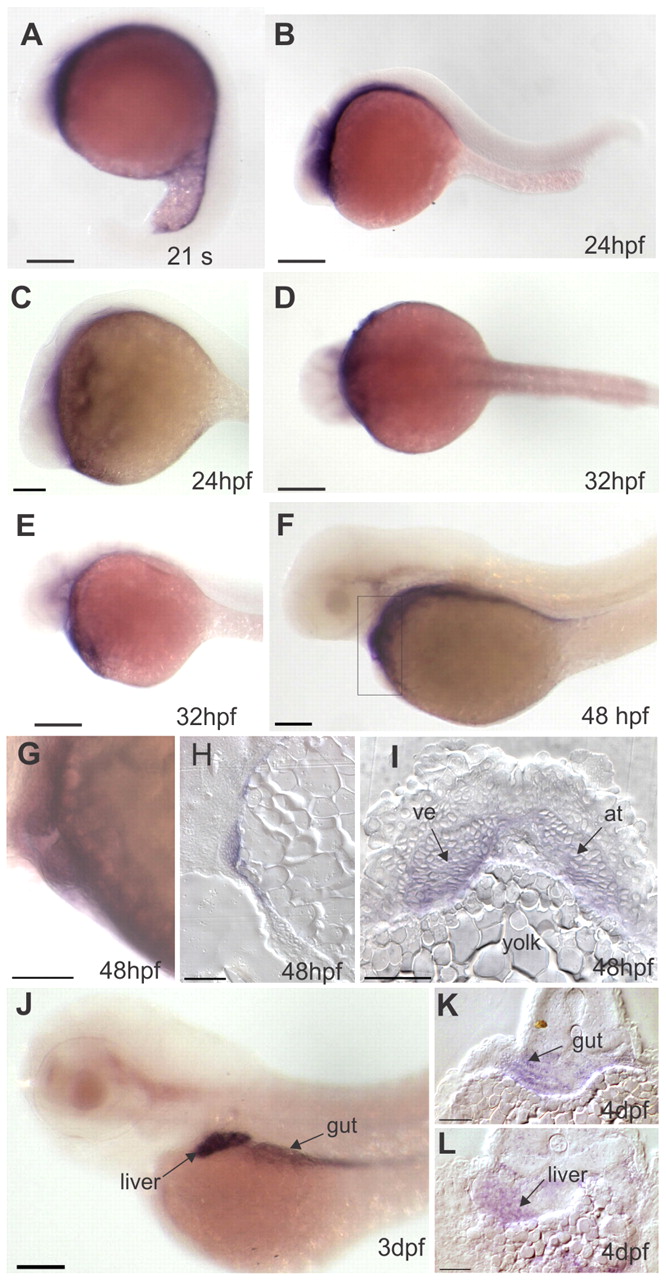
Fig. 2 Expression patterns of bcdo2 during zebrafish development. (A-C,E-G,K) Lateral views, anterior is leftwards. (D) Dorsal view (anterior is leftwards; (H) sagittal section; (I,K,L) transverse cross-sections. (A) During late segmentation stages, bcdo2 is expressed in ventral cell layers. (B-E) At 24 to 32 hpf, bcdo2 expression is found in anterior parts adjacent to the yolk. (F,G) At 48 hpf, bcdo2 is expressed in cardiac and pericardial cells, as well as in endodermal tissues. In G, a higher magnification of the developing heart is shown. (H,I) Sagittal and transverse cross-sections though the cardiac region reveal expression of bcdo2 both in the atrium and the ventricle. (J) At early larval stages, bcdo2 is expressed in cells of the (K) gut and (L) liver. Scale bars: 100 μm in A-D,G,J; 50 μm in E,F,H,I,K,L. at, atrium; ve, ventricle.